7A.1 - Organic Mass Spectrometry
A mass spectrum reveals the mass of a whole molecule and the charged fragments produced when it breaks apart. Use both kinds of evidence to suggest organic structures, compare candidates and distinguish a supported suggestion from a unique identification.
From Spectrum To Evidence
A liquid product has been prepared and purified, but a boiling temperature alone is not enough to identify it. A mass spectrum can give two kinds of evidence:
- the mass of the whole molecule, from the molecular ion peak
- possible pieces of the molecule, from fragment peaks
[DIAGRAM: asset_name: Simplified Organic Mass Spectrum; asset_slug: edexcel_a_level_chemistry_l040_simplified_organic_mass_spectrum; recommended_method: deterministic_chart; description: Simplified organic mass spectrum with peaks at m/z 15, 29, 31, 45 and 60, the molecular ion peak at m/z 60 gives relative molecular mass 60 and the base peak at m/z 31 is the most abundant fragment.]
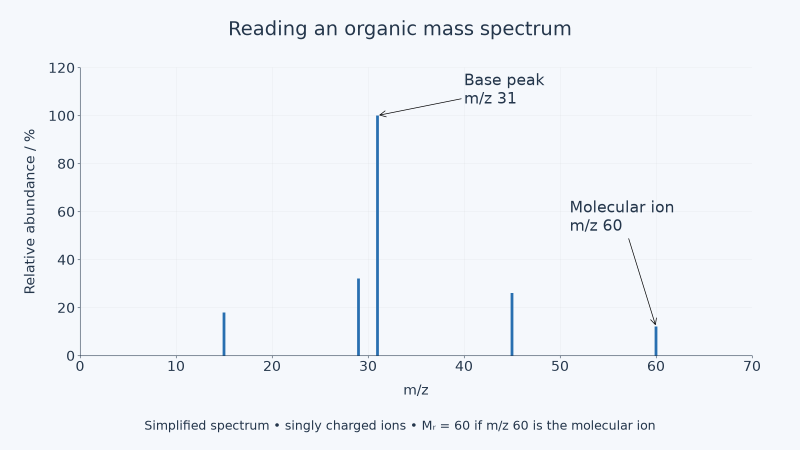
The same simplified spectrum can be read as data:
| m/z | Relative abundance / % | First useful reading |
|---|---|---|
| 15 | 18 | a small fragment ion, often in simple organic spectra |
| 29 | 32 | a fragment ion, often in simple hydrocarbon chains |
| 31 | 100 | the base peak, the most abundant ion in this spectrum |
| 45 | 26 | another fragment ion |
| 60 | 12 | the molecular ion peak, so for a singly charged molecular ion |
Base peak
The tallest peak in a mass spectrum: the most abundant ion reaching the detector. It is often a stable fragment, not the whole molecule.
Molecular ion peak
The peak () produced by the whole molecule after it loses one electron. Its value gives the relative molecular mass when its charge is +1.
Tallest is not heaviest
The peak at is the base peak, the most abundant fragment ion. The molecular ion peak is at , so for a singly charged ion , not 31.
The Molecular Ion Peak
In a mass spectrometer, an organic molecule can lose an electron to form a positive molecular ion. The molecular ion contains the whole molecule, so its relative mass is the relative molecular mass of the compound.
For the spectra used at this level, treat the molecular ion as singly charged:
Relative molecular mass from a mass spectrum
Condition: the molecular ion carries a charge. has no unit; is a mass-to-charge ratio.
So if the molecular ion peak is at , then:
Relative molecular mass has no unit. The axis is a mass-to-charge ratio, and the numerical equality works here because the ion has charge .
Do not automatically choose the rightmost peak. Small isotope peaks such as M+1 can lie beyond the molecular ion, and some molecular ions are weak or absent. Use the labelled molecular ion or the supplied identification evidence.
Worked example: determining Mr
A simple organic compound gives major peaks at , , and a smaller molecular ion peak at .
Determine the relative molecular mass of the compound.
Route:
- Identify the molecular ion peak, not the base peak.
- Use the value of that peak.
- State the relative molecular mass.
The molecular ion peak is at . For a singly charged molecular ion, the relative molecular mass is therefore .
The smaller height of the molecular ion peak does not make it less useful. It still represents the whole molecule.
Common-error contrast
Tempting but wrong: "The tallest peak gives ."
Correct: the tallest peak is the base peak. It may be a very stable fragment ion. Use the molecular ion peak for .
Why Fragment Peaks Form
The molecular ion has excess energy. It can break into smaller pieces, and if one of those pieces keeps the positive charge it can be detected as a fragment ion.
The neutral piece is not detected by the mass spectrometer. This is why a mass spectrum records charged fragments, not every piece formed in the break-up.
For a molecule represented as , fragmentation can be shown in a simplified way:
The molecular ion is usually a radical cation: it has both a positive charge and an unpaired electron. The neutral radical carries no charge. Simplified exam equations may omit the radical dot; conserve atoms and total charge in either notation.
Only gives a peak. If the positive charge stayed on instead, then would give the peak.
Useful fragment masses
You do not need to memorise a huge library of fragments, but you should be able to calculate and use common ones.
| Fragment ion | Mass calculation | m/z for a ion | What it can suggest |
|---|---|---|---|
| 15 | a methyl group is present, common in many organic molecules | ||
| 29 | an ethyl-sized fragment | ||
| 31 | a part, often useful for primary alcohols | ||
| 43 | a propyl-sized fragment, but not unique | ||
| 45 | an oxygen-containing two-carbon fragment |
Continue this lesson.
You’re previewing NovaLearn without an account. Create a free account to read the rest.
Create free account