3.8.4.3 - Genetic fingerprinting
Genetic fingerprinting compares DNA between samples. Some parts of the genome vary a lot between individuals, so amplifying, separating and visualising those regions gives a pattern of bands that is very unlikely to be identical in two unrelated people.
VNTRs and the odds of a chance match
Two people's DNA can be told apart by counting repeats: one short sequence, lined up a different number of times.
Picture the sequence GATA repeated 8 times in a row at one place on a chromosome, and 12 times at the same place in someone else. Stretches like this are called variable number tandem repeats, or VNTRs, and an organism's genome contains many of them.
Variable Number Tandem Repeats (VNTRs)
Short, repeated DNA sequences found at particular loci in the genome, where the number of repeats varies between individuals.
How repeat number sets fragment length
More repeats make a longer stretch of DNA, so the person with 12 copies has a longer fragment containing that locus than the person with 8.
Genetic fingerprinting
A technique used to compare DNA samples by analysing the lengths of DNA fragments containing variable repeated sequences such as VNTRs.
Many VNTR regions used for fingerprinting are in non-coding DNA, so differences in repeat number often have little or no direct effect on phenotype. Natural selection therefore does not strongly remove this variation, and a population can hold many versions of each locus.
VNTR alleles are inherited from parents. Here an allele means one version of the locus, with a particular number of repeats. At each autosomal locus a child inherits one allele from each biological parent. Two different-length alleles usually give two bands at that locus, while one visible band can represent two equal-length alleles.
Why a full match is very unlikely
A single locus is not enough for confident identification, because two people could happen to share the same fragment length there. The power of the method comes from comparing several loci.
Suppose unrelated people share a given fragment length at one locus 1 time in 10. If five loci are inherited independently, the chance of matching at all five is . These numbers are made up, but the idea holds: each extra locus multiplies the chance by another fraction, so it falls quickly.
Close relatives share more alleles than unrelated people, so their profiles are more alike.
Watch out
Don't write that every person has unique VNTRs. Unrelated people can share a repeat number at one locus, and identical twins usually share a whole profile. Write that the probability of two individuals having the same VNTRs is very low, because many loci are compared.
Even a full match has a limitation: it compares fragment lengths at the chosen loci, so it does not reveal every base in the fragment or the rest of the genome.
How a genetic fingerprint is produced
Five steps turn a trace of DNA into a pattern of bands whose positions reflect repeat number.
The PCR-based route
- Extract DNA from a suitable biological sample, such as blood containing white blood cells, saliva, a hair root or plant tissue. Label samples and avoid cross-contamination, because PCR would copy stray DNA too.
- Amplify selected repeat regions with PCR. A crime-scene sample may contain too few copies of the target DNA to give a detectable, complete profile. Primers bind to known sequences flanking each repeat region, so each copy contains the whole repeat region and more repeats give a longer fragment. Repeated PCR cycles (3.8.4.1) then build up many copies from the small starting sample. PCR is an in vitro way of cloning DNA, which is why AQA describes these as fragments ‘cloned by PCR’. It copies the individual's repeat-number variation; it does not create it.
- Separate the products by electrophoresis. Load products and a size ladder (DNA fragments of known length) into separate wells in a gel near the negative electrode. DNA has a negative charge from phosphate groups, so it moves towards the positive electrode, the anode. The gel holds back longer fragments more strongly, so shorter linear fragments travel further during the same run.
- Detect the DNA. A DNA stain, or a fluorescent label carried on the primers into the PCR products, makes separated fragments detectable. A band represents many molecules of a similar length, not a single DNA molecule.
- Compare corresponding loci. Use known fragment sizes and the same analysis conditions. Compare fragment lengths and inheritance patterns rather than brightness, which depends largely on how much DNA is in a band.
The chain is repeat-number variation → different fragment lengths → different migration distances → a pattern that can be compared.
The restriction-and-probe route
An older route reaches the same chain without PCR. Restriction endonucleases cut extracted DNA at recognition sequences flanking repeat regions, so different repeat numbers give different fragment lengths.
A genome gives many restriction fragments, so probes pick out the relevant ones. After electrophoresis, DNA is denatured and transferred to a membrane, and labelled probes hybridise to complementary repeat-region sequences. Washing removes unbound probe, and detecting the labels reveals the selected bands.
Targeted PCR products can be sized directly, so restriction digestion and membrane probing are not compulsory after PCR. If a question supplies a particular method, explain that method rather than adding steps.
Reading gels for matches and parentage
Band position is the evidence on a gel: it gives you fragment length, and length is what you compare between samples.
Reading band positions
Find the wells, the direction of migration and the labelled sample lanes first, then apply these rules:
- Bands that are level with each other in the same gel represent fragments of the same estimated length.
- Bands further from the wells in the direction of migration are shorter, and longer fragments remain closer to the wells. The page could show a gel in any orientation.
- A mismatch in position means a different fragment length. For example, an allele with a 3-base deletion has fewer bases, so its band travels further.
- Assignment to a locus requires a labelled locus or the assay design: four bands do not automatically mean four loci.
Exam tip
Asked how to find fragment sizes, give both steps: run fragments of known length, then compare their positions with the unknown bands. Describing electrophoresis alone, or stopping at the first step, can lose marks.
A crime-scene gel
A gel gives the following band positions, measured from the wells:
| Sample | Band positions / mm |
|---|---|
| Crime scene sample | 14, 29, 47, 63 |
| Suspect A | 14, 29, 47, 63 |
| Suspect B | 14, 33, 47, 63 |
In the figure the wells are at the top, beside the negative electrode. Compare each band in the Scene lane with the level position in each suspect's lane.
[DIAGRAM: asset_name: 3.8.4.3-worked-gel; asset_slug: 3.8.4.3-worked-gel; recommended_method: retained_png; caption: Suspect A matches all four detected scene fragments, Suspect B has a different band at 33 mm.; description: Suspect A matches all four detected scene fragments; Suspect B has a different band at 33 mm.]
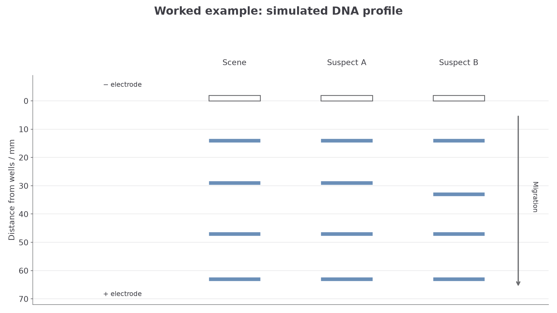
Simulated data. The ruler measures migration distance, not fragment length in base pairs.
A strong exam answer reasons like this:
- The crime-scene sample and Suspect A have bands at the same four positions, so all four detected fragments have matching lengths. The table does not specify how many loci produced them.
- Suspect B differs at one band, because 33 mm is not the same position as 29 mm. So Suspect A is the closer match.
- Even a complete match does not by itself prove guilt. The DNA could have come from Suspect A, but the sample could have been deposited innocently, transferred indirectly or contaminated.
Testing a possible parent
Compare the same locus in all family members. Each child allele must be accounted for by a parental allele, with one inherited from each parent. A band pattern alone cannot assign parent of origin when both parents share an allele.
If the mother carries both of the child's alleles at a locus, either could be the paternal one, so that locus excludes only a candidate father who has neither.
Continue this lesson.
You’re previewing NovaLearn without an account. Create a free account to read the rest.
Create free account