3.3.2 - Gas exchange
An amoeba needs no lungs, an insect pipes air straight to its muscles, and a fish takes oxygen from water. All exchange gases by diffusion, and three features keep that fast: a large surface area, a short diffusion pathway and a maintained concentration gradient. Ventilation helps maintain the gradient by moving the air or water at the surface. This lesson compares these systems, then covers human breathing, lung disease data and how gas exchange is observed and measured.
Gas exchange in single-celled organisms
A single-celled organism exchanges gases across its whole surface, so it needs no lungs, gills or tracheae.
An amoeba’s cell-surface membrane has all three features of a good exchange surface:
- Large surface area: a tiny cell has a large surface area to volume ratio, as you saw in 3.3.1.
- Short diffusion pathway: no part of the cell is far from the surface.
- Maintained concentration gradients: respiration uses oxygen and produces carbon dioxide continuously, which keeps oxygen lower and carbon dioxide higher inside the cell than outside.
As organisms get larger, their surface area to volume ratio falls and their inner cells lie further from the surface, so simple diffusion across the body surface becomes too slow. Larger organisms therefore have specialised exchange surfaces.
Gas exchange in insects
Insects deliver air directly to their tissues through tubes, rather than carrying oxygen in the blood as mammals do.
From spiracle to muscle cell
The tracheal system has three main parts:
- Spiracles: small openings on the body surface.
- Tracheae: larger air-filled tubes. Chitin strengthens their walls, so they do not collapse.
- Tracheoles: very fine branches that run between cells, giving a short diffusion pathway to respiring tissues.
Trace that route across the diagram, from the spiracle on the left to the muscle cells on the right. Then compare the arrows: oxygen moves in and carbon dioxide moves out, in opposite directions.
[DIAGRAM: asset_name: 3.2 - Gas exchange - Diagram 1; asset_slug: 3.2 - Gas exchange - Diagram 1; recommended_method: retained_png; caption: An insect spiracle opens into branching tracheae and fine tracheoles close to muscle cells, oxygen and carbon dioxide move in opposite directions.; description: An insect spiracle opens into branching tracheae and fine tracheoles close to muscle cells; oxygen and carbon dioxide move in opposite directions.]
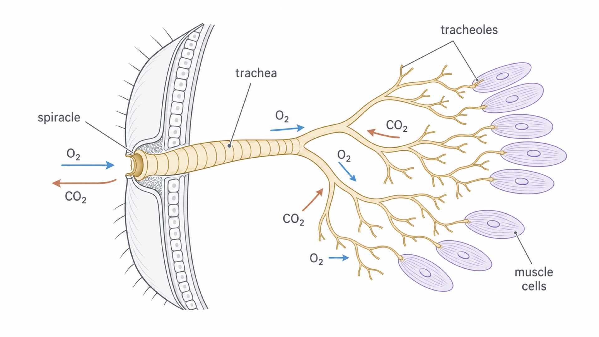
The key detail is on the far right: tracheoles end right beside the muscle cells. That very short final pathway makes rapid gas exchange possible without blood transport.
Gases move through this system in three ways:
- Diffusion: respiring cells use oxygen and produce carbon dioxide, so oxygen diffuses along the tubes towards the cells and carbon dioxide diffuses out.
- Ventilation: during activity, abdominal muscle contractions can pump the system, creating a mass movement of air.
- Water withdrawal: the ends of the tracheoles normally hold a little watery fluid, and oxygen diffuses slowly through liquid. During intense activity, soluble products of respiration lower the water potential of muscle cells, so water moves from the tracheole ends into these cells by osmosis. Air then extends further along the tracheoles, shortening the slow path through liquid.
Diffusion is fast only over short distances, which is one reason insects cannot grow to an unlimited size.
Balancing gas exchange and water loss
Efficient gas exchange requires a permeable surface, but a permeable surface also loses water, which is a problem on land. Insects reduce the loss in three ways:
- a waterproof cuticle covers the body surface
- spiracles can close, so they need not stay open when oxygen demand is low
- the exchange surfaces lie inside the body, not exposed to dry air
This is a structural and functional compromise. If spiracles stay closed, water loss falls, but gas exchange is reduced. If spiracles stay open, oxygen supply improves, but evaporation increases.
Gas exchange in fish
Gills give a fish a huge exchange surface, and opposite flows keep oxygen diffusing along the whole of it.
Gill structure and ventilation
For most bony fish, diffusion across the body surface cannot meet the demands of their volume of respiring tissue. Gills provide a much larger, thin surface in contact with water.
Each gill is supported by a gill arch. Extending from the arch are many gill filaments, and at right angles to each filament are many thin gill lamellae, the main gas exchange surfaces. Together they provide:
- a very large surface area, from many lamellae on many filaments
- a short diffusion pathway across a thin epithelium
- a rich blood supply in capillaries close to the water, carrying oxygen away
Ventilation brings fresh water over the gills in a continuous one-way stream. The mouth opens and the floor of the buccal (mouth) cavity lowers, reducing the pressure inside and drawing water in. The mouth then closes and the floor rises, so higher pressure pushes water across the gills and out beneath the operculum (gill cover).
How countercurrent flow keeps the gradient
In countercurrent flow, blood in the lamellae flows in the opposite direction to the water passing over them. On the left of the diagram, find the arch, filaments and lamellae. Then, on the right, follow both arrows along one lamella.
[DIAGRAM: asset_name: fish-countercurrent-v2; asset_slug: fish-countercurrent-v2; recommended_method: retained_png; caption: Opposite water and blood flow maintains an oxygen gradient at every position across a fish gill lamella.; description: Opposite water and blood flow maintains an oxygen gradient at every position across a fish gill lamella.]
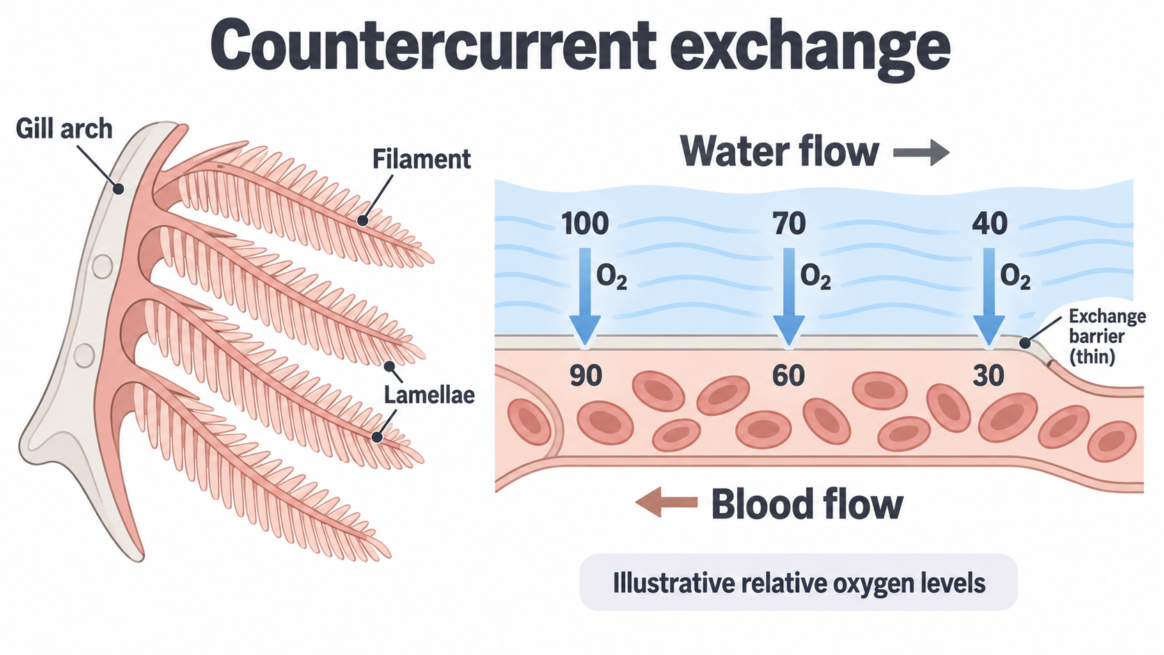
Read the oxygen levels by position: water 100 is beside blood 90, water 70 beside blood 60, and water 40 beside blood 30. At every position the water holds more oxygen than the blood beside it, so oxygen diffuses into the blood. These are illustrative relative levels, not measured concentrations or percentages.
Follow the blood from right to left: it gains oxygen, rising from 30 to 90. As it does, it keeps moving towards fresher water, so the gradient is maintained along the whole lamella and equilibrium is not reached.
Now compare parallel flow, where blood and water move in the same direction. Their oxygen levels converge as exchange proceeds, so the gradient and net diffusion decline. At equilibrium there is no net diffusion. Countercurrent flow therefore allows much more oxygen to be absorbed.
Gas exchange in dicotyledonous plants
A leaf both photosynthesises and respires, so the direction of its gas exchange changes with the light.
Gas routes through the leaf
- In daylight, photosynthesis usually exceeds respiration, so overall the leaf takes in and releases . Photosynthesis uses faster than respiration makes it, so its concentration in the air spaces falls below that of the outside air and diffuses in.
- In darkness, photosynthesis stops and respiration continues, so the leaf takes in and releases .
On the leaf section, start at the stoma in the lower epidermis and move up into the spongy mesophyll. That is the main route for gases into and out of the leaf.
[DIAGRAM: asset_name: 3.2 - Gas exchange - Diagram 3; asset_slug: 3.2 - Gas exchange - Diagram 3; recommended_method: retained_png; caption: A dicot leaf section shows mesophyll air spaces linked to the outside through a stoma controlled by guard cells.; description: A dicot leaf section shows mesophyll air spaces linked to the outside through a stoma controlled by guard cells.]
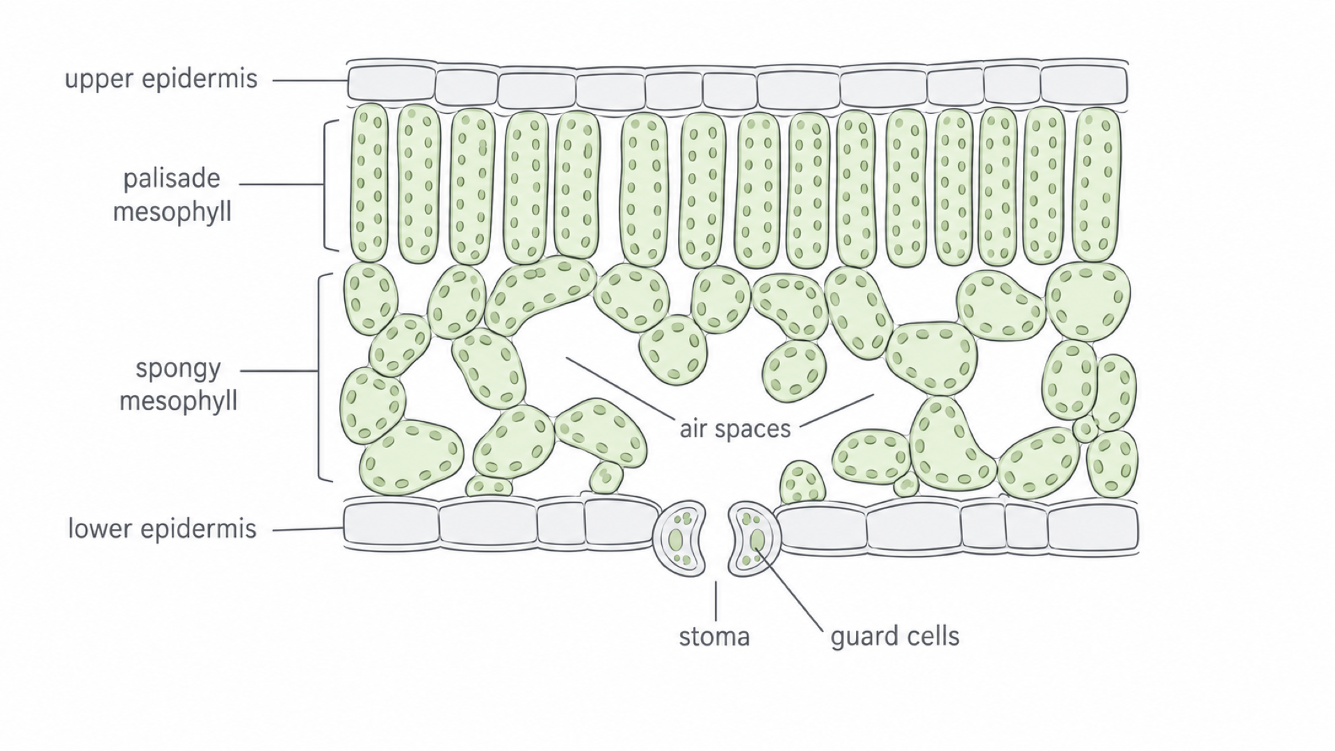
The leaf is adapted for gas exchange in these ways:
- It has many stomata, especially in the lower epidermis, so mesophyll cells are not far from a route to the outside air.
- Large interconnecting air spaces in the spongy mesophyll keep the diffusion pathway short, because gases move through air, not through a thick layer of tissue.
- Many mesophyll cells are exposed to those air spaces, creating a large internal surface area.
- Guard cells open and close the stomata, controlling gas exchange and water loss.
Xerophytes and water loss
Open stomata improve carbon dioxide uptake, but they also allow water vapour to diffuse out. This is the same kind of compromise seen in insects. Xerophytes, such as marram grass, are plants adapted to reduce this water loss.
| Adaptation | How it helps reduce water loss |
|---|---|
| Thick waxy cuticle | Reduces evaporation from the leaf surface |
| Rolled leaves | Trap humid air and reduce the water-vapour concentration gradient |
| Hairy leaves | Trap still, moist air near the surface |
| Sunken stomata | Keep stomata in a sheltered, humid pocket |
| Reduced leaf surface area | Reduces the area from which water can be lost |
Humid air near stomata reduces the water-vapour concentration gradient to the outside. You can also describe this as a smaller water potential gradient, because humid air has a higher water potential than dry air. Sheltering also stops wind removing this humid layer so quickly.
Limiting water loss has a cost: closed stomata and restricted gas movement can reduce carbon dioxide entry and photosynthesis.
A full answer follows a chain: where air is trapped, why it becomes humid, why the gradient falls, and why less water vapour diffuses out.
The human gas exchange system
Air travels down ever narrower tubes to the alveoli, where only two thin cells separate it from the blood.
From trachea to alveoli
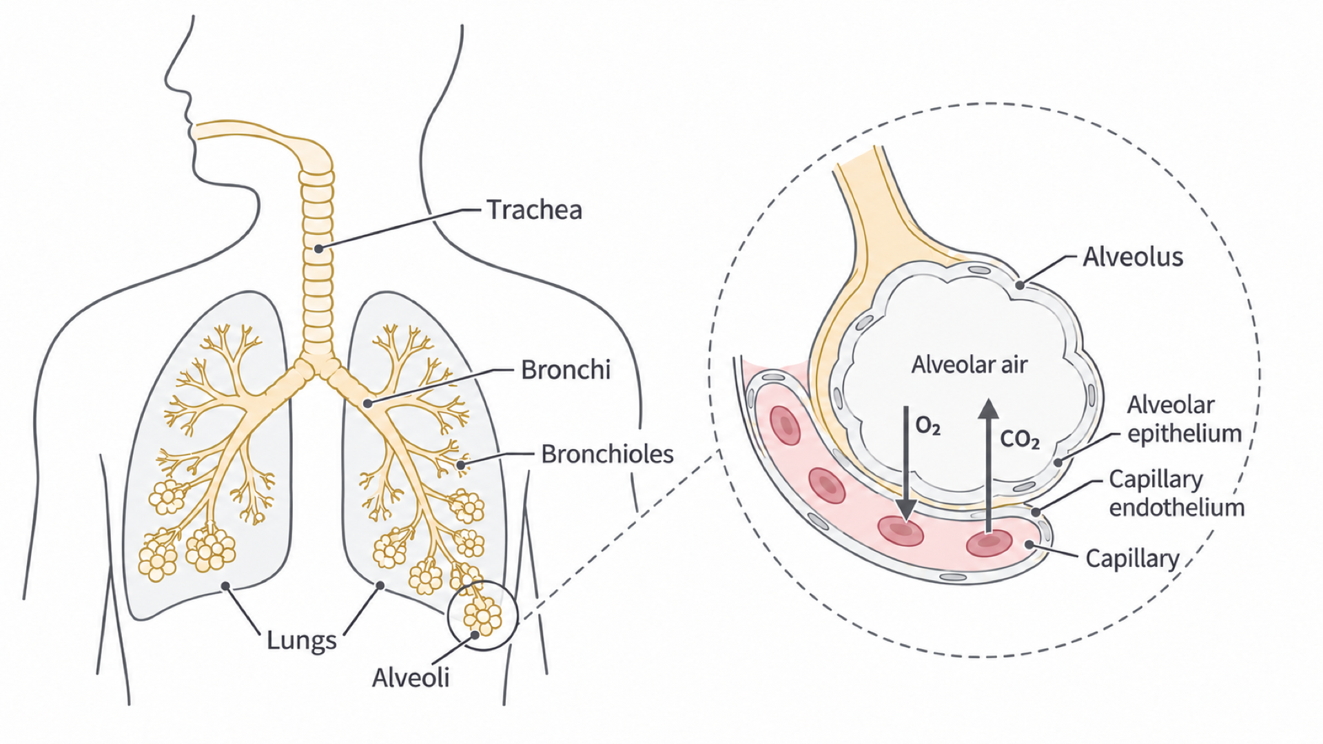
Air enters the two lungs through the trachea, then passes along bronchi and bronchioles into the alveoli. Trace that pathway on the left of the diagram, then use the inset to see the exchange surface up close.
[DIAGRAM: asset_name: 3.2 - Gas exchange - Diagram 4; asset_slug: 3.2 - Gas exchange - Diagram 4; recommended_method: retained_png; caption: Air passes from trachea to bronchi, bronchioles and alveoli, alveoli lie beside blood capillaries.; description: Air passes from trachea to bronchi, bronchioles and alveoli; alveoli lie beside blood capillaries.]
- Trachea: held open by C-shaped rings of cartilage; lined with ciliated epithelium and goblet cells.
- Bronchi: similar to the trachea, but narrower and with less cartilage as they branch.
- Bronchioles: narrow airways with smooth muscle and elastic tissue that help control airflow.
- Alveoli: tiny air sacs that form the main gas exchange surface.
Goblet cells secrete mucus, which traps dust and microorganisms. Cilia then move the mucus up towards the throat, where it can be swallowed. This helps protect the delicate alveoli from infection and particle damage.
The alveolar exchange surface
In the inset, oxygen diffuses from the air, across the alveolar epithelium and the capillary endothelium, into the blood. Carbon dioxide diffuses the opposite way, from the blood into the alveolar air. The same three features make this exchange fast:
- Large surface area: there are many millions of alveoli.
- Short diffusion pathway: the alveolar epithelium is a single layer of squamous (flattened) epithelial cells, and the capillary endothelium is also one cell thick. Narrow capillaries place red blood cells close to the alveolar wall, shortening the path from alveolar air to haemoglobin.
- Maintained gradients: ventilation keeps a high concentration and a low concentration in alveolar air. Blood flow carries oxygen away and brings carbon dioxide to the alveoli.
Watch out
Don’t write that the cells have “thin membranes”. Say that the alveolar epithelium and the capillary endothelium are each only one cell thick, so the diffusion pathway is short.
Ventilation and breathing
Muscles change the volume of the thoracic cavity, pressure falls or rises as a result, and air moves.
Ventilation is the movement of air into and out of the lungs. It keeps fresh air reaching the alveoli, which helps maintain concentration gradients for gas exchange.
Muscles, volume and pressure
Two physical ideas explain ventilation:
- when the volume of a gas in a closed space increases, its pressure decreases
- gases move from higher pressure to lower pressure
The diaphragm and the intercostal muscles between the ribs change the volume. The external and internal intercostals act antagonistically: they pull the ribs in opposite directions, so when one set contracts, the other relaxes. The external intercostals raise the ribs, and the internal intercostals lower them.
Compare the two panels: the wider ribcage and flatter diaphragm in inspiration, then the narrower ribcage and domed diaphragm in expiration at rest.
Continue this lesson.
You’re previewing NovaLearn without an account. Create a free account to read the rest.
Create free account