3.6.2.2 - Synaptic transmission
An action potential cannot cross a chemical synapse, so a chemical neurotransmitter carries its effect over the gap. Use that step to explain one-way transmission, summation, inhibition, muscle stimulation and drug effects.
Structure of a synapse
A synapse converts an action potential into a chemical step and back; its structure shows how.
A chemical synapse is a junction where one cell releases a neurotransmitter that changes the activity of another cell. A narrow extracellular gap, the synaptic cleft, separates the cells, so neurotransmitter has only a short distance to diffuse.
Work down the diagram from the presynaptic neurone.
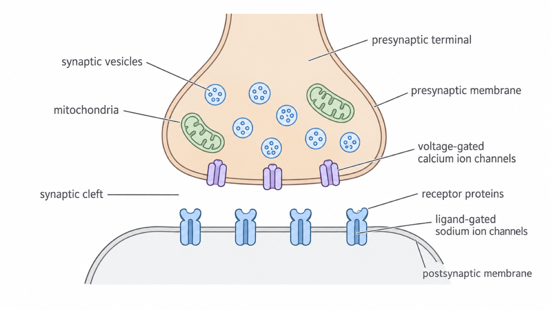
The presynaptic side
The presynaptic terminal, or synaptic knob, is the swollen end of the axon.
- Vesicles are membrane-bound sacs of neurotransmitter. A vesicle is the container, not the neurotransmitter itself. It can fuse with the presynaptic membrane, releasing its contents by exocytosis.
- Voltage-gated calcium channels in the presynaptic membrane open when an action potential arrives, linking its arrival to release.
- Mitochondria supply ATP for ion pumps and for recycling vesicles and transmitter.
The postsynaptic side
The postsynaptic membrane contains receptors with binding sites complementary to the transmitter, facing the cleft.
In this diagram, each blue protein does two things: the cup is the receptor and the pore below is a ligand-gated sodium ion channel, so binding opens the channel directly. A ligand-gated channel responds to a binding molecule; a voltage-gated channel responds to membrane potential.
Transmission across a cholinergic synapse
Seven linked steps pass excitation across a cholinergic synapse, then reset it for the next impulse.
A cholinergic synapse uses acetylcholine, abbreviated ACh, as its neurotransmitter.
- An action potential reaches the presynaptic terminal and depolarises its membrane.
- Voltage-gated calcium channels open, so calcium ions enter from the cleft by facilitated diffusion, down their electrochemical gradient.
- Calcium entry triggers vesicles to fuse with the presynaptic membrane, releasing acetylcholine by exocytosis.
- Acetylcholine diffuses across the cleft and binds to complementary receptors on the postsynaptic membrane.
- Ligand-gated channels open. Net sodium-ion entry makes the inside less negative, producing an excitatory postsynaptic potential. If this brings the trigger region to threshold, a new action potential is generated there.
- Acetylcholinesterase, an enzyme in the cleft, hydrolyses acetylcholine into choline and ethanoate (often described as ethanoic acid). Receptors stop being activated and the channels close, so stimulation does not persist and each successive impulse needs newly released acetylcholine.
- Choline is taken back into the presynaptic terminal and used to make new acetylcholine, packaged in vesicles. ATP from mitochondria supports this resynthesis.
Watch out
Acetylcholine crosses the cleft, but the vesicles stay in the terminal and the action potential does not diffuse across. Binding opens sodium channels, not calcium channels: calcium entry happens earlier, in the presynaptic terminal.
A postsynaptic potential is graded: its size depends on the input. It may be too small to trigger an action potential, whose size is all-or-nothing. This difference is the basis of summation.
Unidirectionality, summation and inhibition
Why does transmission run only one way, and why can one input fail where three succeed?
Why transmission is one-way
Transmission at a chemical synapse is unidirectional: it goes only from the presynaptic cell to the postsynaptic cell. Vesicles are found only in the presynaptic terminal and receptors only on the postsynaptic membrane, so transmission cannot run the other way.
In an axon the reason is different: the refractory region behind an impulse stops it running backwards.
Adding inputs together
One impulse may release too little acetylcholine to open enough channels, so the depolarisation stays below threshold. If postsynaptic potentials overlap, more ligand-gated channels are open at once, more sodium ions enter and the depolarisation is larger.
Temporal and spatial summation
Temporal summation occurs when repeated impulses arrive from the same presynaptic neurone close enough together that their postsynaptic potentials overlap before the previous depolarisation has decayed.
Spatial summation occurs when inputs from different presynaptic neurones produce overlapping postsynaptic potentials in the same postsynaptic neurone.
Either way, the combined depolarisation may reach threshold even if each input alone is insufficient. It is the postsynaptic potentials that sum; full action potentials do not fuse into one larger action potential.
Inhibitory synapses
At an inhibitory synapse, receptor activation opens chloride channels, letting chloride ions in, or potassium channels, letting potassium ions out, depending on the receptor. Either way the inside usually becomes more negative: hyperpolarisation. The membrane is now further from threshold, so more sodium ions must enter to reach it and firing becomes less likely.
Exam tip
Give both halves: the inside is more negative, so more sodium ions must enter to reach threshold. On AQA's 2024 question only about 18% earned both marks.
Excitatory and inhibitory effects combine, so add them with their signs.
Worked example: adding postsynaptic potentials
A simplified model with no decay has a resting potential of −70 mV and a threshold of −55 mV. Each excitatory input adds +6 mV and an inhibitory input adds −5 mV. Test two excitatory inputs, three, and three plus one inhibitory input.
Plan: Add each input to the resting potential with its sign, then compare with threshold.
- Two: −70 + (2 × 6) = −58 mV, below threshold.
- Three: −70 + (3 × 6) = −52 mV, above threshold.
- Three plus inhibition: −52 − 5 = −57 mV, below threshold.
Answer: Only three excitatory inputs without inhibition fire the neurone.
Check: Threshold is 15 mV above rest: 18 mV clears it, but 12 mV and 13 mV do not.
The limitation of this model is that real postsynaptic potentials decay, and their size at the trigger region depends on timing and distance from it.
Continue this lesson.
You’re previewing NovaLearn without an account. Create a free account to read the rest.
Create free account