3.1.4.1 - Protein structure
Haemoglobin dissolves and carries oxygen, while collagen forms rope-like fibres, yet both are chains drawn from the same set of twenty amino acids. Follow how sequence becomes shape and shape becomes function, then detect proteins with the Biuret test and separate amino acids by chromatography.
Amino acids
Amino acids are the monomers from which proteins are made, and the twenty common to all organisms share one layout.
Start at the central carbon atom in the diagram, then follow its four bonds: to an amine group (NH₂), a carboxyl group (COOH), a hydrogen atom and an R group, or side chain.
[DIAGRAM: asset_name: 1.4.1 - General properties of proteins - Diagram 1; asset_slug: 1.4.1 - General properties of proteins - Diagram 1; recommended_method: retained_png; caption: General structure of an amino acid showing the central carbon bonded to NH2, COOH, H, and R, with each group labelled.; description: General structure of an amino acid showing the central carbon bonded to NH2, COOH, H, and R, with each group labelled.]
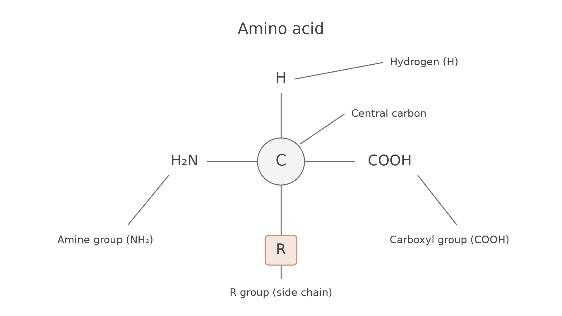
The first three are the same in every amino acid, and only the R group differs. The shared groups let any two amino acids join in the same way. The R group gives each one its own chemistry:
- Some R groups carry a charge at a given pH.
- Some are uncharged but polar: they have slightly positive and slightly negative regions, so they attract water.
- Some are non-polar, so water attracts them only weakly, as with the fatty acid tails in 3.1.3.
Glycine's R group is just H, alanine's is CH₃ and cysteine's contains sulfur. These differences decide how a chain folds, and therefore what a protein can do.
Peptide bonds and polypeptides
Amino acids join by a condensation reaction.
You met this reaction building polysaccharides in 3.1.2 and triglycerides in 3.1.3. Follow the diagram from the top row to the bottom:
- The carboxyl group of one amino acid reacts with the amine group of another.
- The OH from the carboxyl group and one H from the amine group leave as a molecule of water.
- The carbon and nitrogen left behind join by a new covalent bond, the peptide bond.
[DIAGRAM: asset_name: 1.4.1 - General properties of proteins - Diagram 2; asset_slug: 1.4.1 - General properties of proteins - Diagram 2; recommended_method: retained_png; caption: Two amino acids joining by condensation, showing removal of OH from one amino acid and H from the other to make water, and the peptide bond left behind.; description: Two amino acids joining by condensation, showing removal of OH from one amino acid and H from the other to make water, and the peptide bond left behind.]
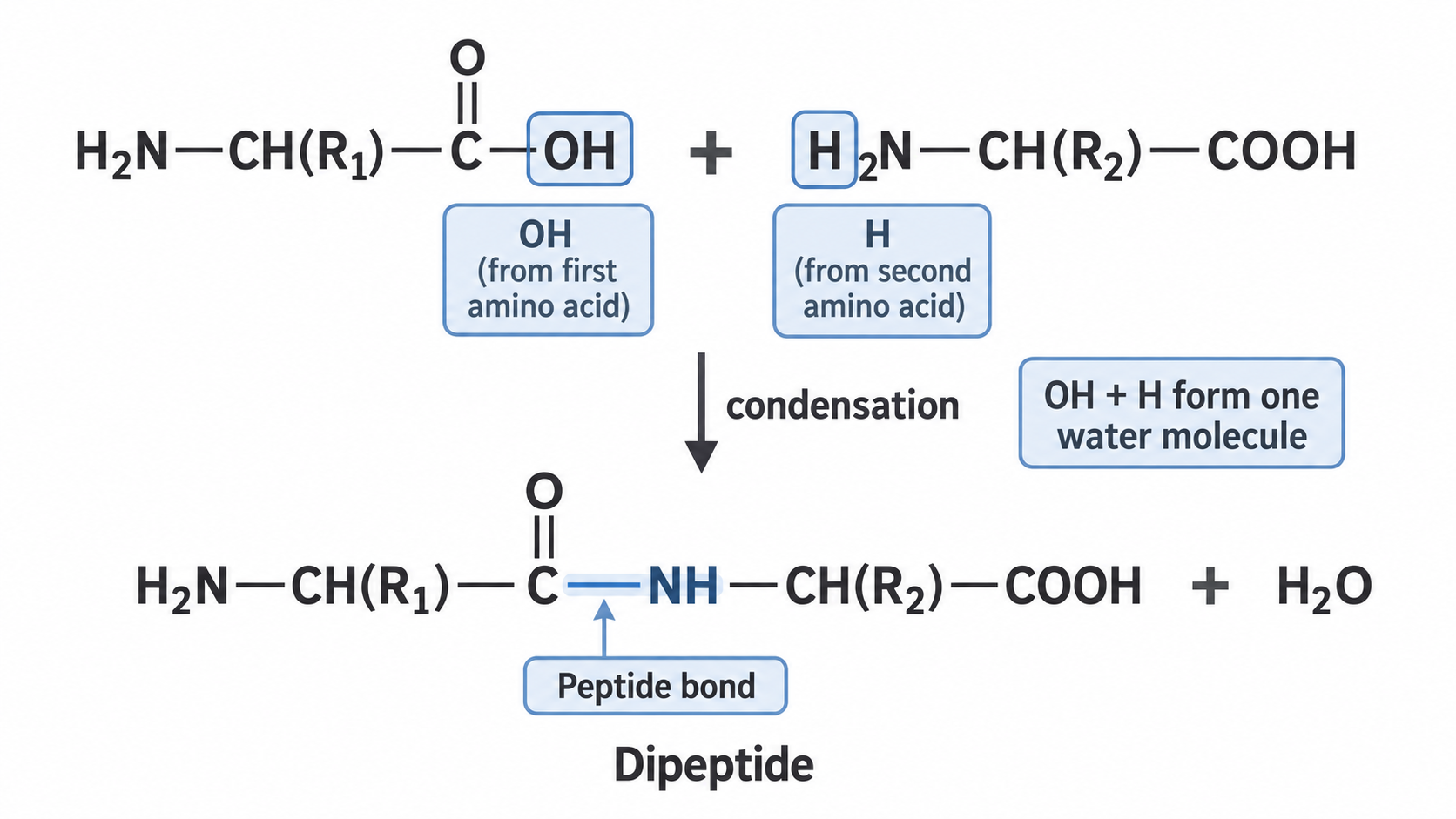
The nitrogen keeps one hydrogen, so the link reads –CO–NH–. The two R groups play no part in it.
The product still has a free amine group at one end and a free carboxyl group at the other, so further condensation reactions can add more amino acids. Each one forms another peptide bond and releases another water molecule. Keep these terms apart:
- A dipeptide is two amino acids joined by a peptide bond.
- A polypeptide is a long chain of many amino acids joined by peptide bonds.
- A functional protein contains one or more polypeptides, folded and, where there are several, joined into the shape it needs to do its job.
Hydrolysis is the reverse: water is added to break peptide bonds. Proteins in food are digested this way before their amino acids can be absorbed.
Levels of protein structure
A polypeptide works only once folded into a specific shape, which builds up in four levels.
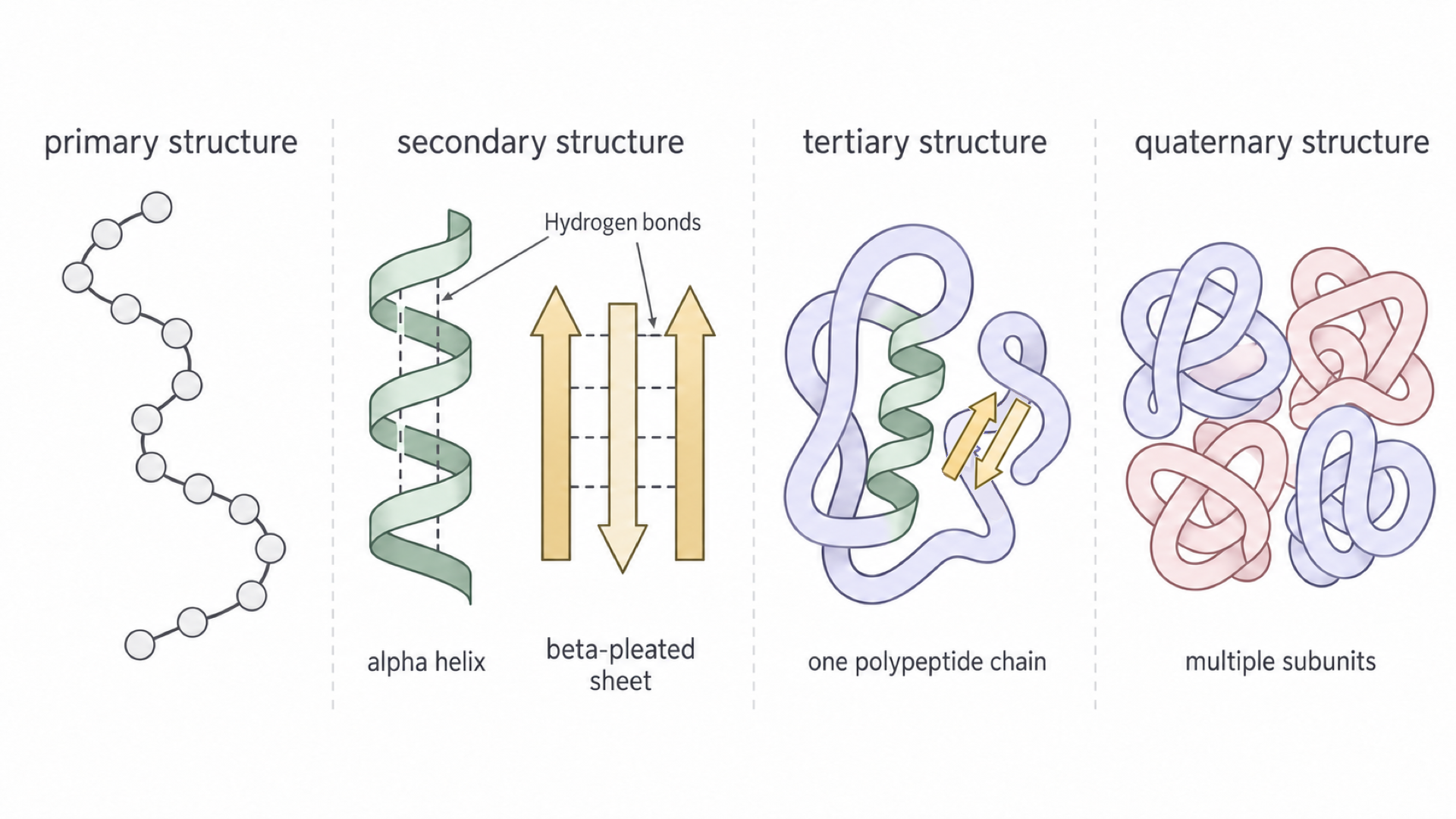
Find each level in the diagram, from left to right, as you read.
[DIAGRAM: asset_name: 1.4.1 - General properties of proteins - Diagram 3; asset_slug: 1.4.1 - General properties of proteins - Diagram 3; recommended_method: retained_png; caption: Protein structure overview showing primary sequence, secondary alpha helix/beta-pleated sheet, tertiary folded polypeptide, and quaternary arrangement of multiple subunits.; description: Protein structure overview showing primary sequence, secondary alpha helix/beta-pleated sheet, tertiary folded polypeptide, and quaternary arrangement of multiple subunits.]
Primary and secondary structure
The primary structure is the sequence of amino acids in a polypeptide chain, joined by peptide bonds. The order matters, not just the number, because it sets where each R group sits and so which bonds can form during folding. Even one changed amino acid can alter the final shape and reduce or change function.
The secondary structure is the regular folding of the chain into an alpha helix or a beta-pleated sheet, held by hydrogen bonds: the dashed lines in the diagram.
These bonds form along the backbone, the repeating N–C–C chain through every amino acid, with R groups sticking out from it. The H of an NH group is slightly positive and the O of a C=O group is slightly negative, so they attract. R groups are not involved, and these are not peptide bonds.
Tertiary and quaternary structure
The tertiary structure is the overall three-dimensional shape of one polypeptide chain. Folding brings amino acids that were far apart in the sequence close together, so their R groups can interact:
| Interaction | What it connects | Significance |
|---|---|---|
| Hydrogen bonds | Suitable polar groups, including R groups | Weak individually, but many together stabilise the fold |
| Ionic bonds | Oppositely charged R groups | Depend on the charges present at the surrounding pH |
| Disulfide bridges | Sulfur atoms from two cysteine R groups | Covalent cross-links that help stabilise the fold |
| Hydrophobic interactions | Non-polar R groups, clustered together because water attracts them only weakly | Give many soluble proteins a non-polar interior |
The quaternary structure is the arrangement of two or more polypeptide chains, or subunits, in a functional protein. Hydrogen bonds, ionic bonds and disulfide bridges also join subunits. Haemoglobin has four subunits and works only when they are assembled correctly. A single-polypeptide protein has no quaternary structure: its tertiary structure is its final shape.
So the levels form a chain: the sequence places the R groups, the R groups set which bonds form, the bonds hold the shape, and the shape decides the function.
How pH and heat undo the shape
Changed conditions can disrupt the bonds holding a protein's shape.
Denaturation
Denaturation is the loss of a protein's functional three-dimensional shape. It does not normally hydrolyse the peptide bonds or change the primary sequence.
A pH change alters hydrogen ion concentration, so R groups can gain or lose hydrogen ions and their charges change. In more acidic conditions, a negative –COO⁻ R group gains a hydrogen ion and loses its charge, so it no longer forms an ionic bond with a positive R group. Some hydrogen bonds break too.
Heating increases molecular motion, which can disrupt the hydrogen and ionic bonds maintaining the fold. Either way, a changed binding site may no longer be complementary to its substrate or other target. Not every small pH or temperature change denatures every protein.
Structure and function
Why does haemoglobin dissolve in red blood cells while collagen forms insoluble fibres?
Both are built from the same set of twenty amino acids. Their sequences differ, so different R groups sit along each chain. The chains then fold and assemble into different shapes, and a protein's function depends on its shape.
Globular and fibrous proteins
Globular proteins fold into compact shapes and are usually soluble, which suits roles such as catalysis and transport. Enzymes and haemoglobin are globular. Polar and charged R groups on the surface attract water, while non-polar R groups are buried inside, so solubility comes from surface chemistry, not from being round.
Haemoglobin has four polypeptide subunits, each containing a haem group with an iron ion that binds oxygen reversibly. Its compact, soluble structure suits transport in red blood cells, and interactions between subunits let binding at one site affect other sites. Lesson 3.3.4.1 develops this.
Fibrous proteins are long, strong and usually insoluble, which suits structural roles. Collagen has three polypeptide chains wound around each other, forming a rope-like molecule. Many of these molecules assemble into fibrils, with covalent cross-links between them, so collagen resists tensile (pulling) forces in tendons and other connective tissues.
| Haemoglobin | Collagen | |
|---|---|---|
| Type | Globular | Fibrous |
| Polypeptides | Four subunits, each with a haem group | Three, wound together |
| Shape | Compact | Long and rope-like, forming cross-linked fibrils |
| In water | Soluble | Insoluble |
| Role | Carries oxygen in red blood cells | Resists pulling forces in tendons |
Functions that depend on binding
Proteins have a wide variety of functions in all living organisms, and many depend on binding another molecule:
- Enzymes catalyse reactions: the active site's shape and chemical groups bind appropriate substrates.
- Antibodies bind antigens: variable regions form binding sites complementary to a particular antigen.
- Channel and carrier proteins move substances across membranes: some regions are compatible with the lipid bilayer and others interact with what they transport.
For any protein named in the specification, explain its role from its own structure, rather than assuming all proteins work by catalysis.
A substitution that changes a charged R group to a non-polar one can change ionic interactions and the way the chain folds or joins other subunits, so a binding site's shape or chemistry changes.
In sickle-cell disease, charged glutamate is replaced by non-polar valine on the surface of haemoglobin's beta chains. Deoxygenated molecules then stick together in long fibres, rather than staying dissolved.
Exam tip
When a changed sequence alters a protein, name the bonds: hydrogen, ionic or disulfide bonds form in different places, so the tertiary structure changes.
A substitution need not alter function: its position and the properties of the replacement matter.
The Biuret test
The Biuret test shows whether a sample contains protein, because it detects peptide bonds.
Copper(II) ions are blue, but in alkaline conditions they bind to peptide bonds and form a purple complex.
- Put the sample in a clean test tube and add sodium hydroxide solution to make the mixture alkaline.
- Add a few drops of dilute copper(II) sulfate solution and mix.
- Look for a colour change. Heating is unnecessary, unlike in the Benedict's test.
Biuret reagent already contains both solutions, so it can replace steps 1 and 2.
- A purple or lilac colour is a positive result: peptide bonds are present, so protein is present.
- A blue colour is a negative result: no protein was detected.
Test a known protein as a positive control and distilled water as a negative control, in separate clean tubes, keeping reagent and sample volumes comparable. Wear eye protection because the alkali is corrosive.
What the colour does not show
The routine test is qualitative, with three limits:
- Purple shows multiple peptide bonds, not which protein is present.
- Free amino acids give no positive result, so fully hydrolysing a protein removes the purple colour.
- Blue means no protein was detected at the sensitivity used. It does not establish zero concentration.
Separating amino acids
In chromatography, different R groups make amino acids travel different distances.
A mixture therefore separates into spots that known standards can identify. You used the same method for sugars in 3.1.2.
The solvent is the mobile phase and the paper is the stationary phase. An amino acid that dissolves better in the solvent and is held less strongly by the paper travels further in the same time.
Running the chromatogram
- Draw a pencil baseline near the bottom of the chromatography paper. Graphite stays on the line, whereas ink would dissolve and move with the solvent.
- Place small spots of each standard and the unknown on the baseline. Dry each spot before adding more sample, so it stays small.
- Stand the paper in a suitable solvent in a covered container, with the solvent below the baseline. Spots in the solvent would dissolve directly into the reservoir.
- Let the solvent rise, then remove the paper and mark the solvent front immediately, before it evaporates.
- Apply a locating reagent such as ninhydrin, because amino acids are colourless, following its handling and heating precautions.
Reading the spots
Measure both distances from the baseline.
Rf value
Both distances use the same unit, so has no unit. With a solvent front at 75 mm and a spot at 30 mm, . Compare with standards developed under the same conditions:
- Two visible spots support at least two separated components.
- One spot need not prove purity, because components may overlap.
- A matching standard suggests identity but is not unique proof.
- A second solvent can resolve spots that travel together in the first.