3.4.2 - DNA and protein synthesis
Muscle and pancreas cells carry the same genes yet make different proteins. Follow a protein-coding gene from DNA through RNA to a polypeptide, then test that explanation with codon tables and experimental data.
Genome and proteome
A muscle cell and an insulin-secreting cell carry the same genes, so why do they make such different proteins?
Genome and proteome
The genome is the complete set of genes in a cell. The proteome is the full range of proteins that a cell is able to produce.
In wider biology, genome usually means the complete genetic material, including non-coding DNA.
Most nucleated body (somatic) cells in one organism inherit essentially the same genome through mitosis. What differs between cell types is gene expression: which genes are switched on and transcribed, and how fast their RNA is translated.
A pancreatic cell and a muscle cell both carry the insulin gene, but the pancreatic cell transcribes it heavily, so it makes and secretes insulin. Different genes expressed means different mRNA, different proteins and a specialised function.
The proteins present also change with developmental stage and conditions, so a change in the proteins a cell is making does not, by itself, show that DNA has mutated. A cell in a new condition may simply express more of an existing enzyme.
Structure of mRNA and tRNA
mRNA carries a gene's code to the ribosome and tRNA delivers the amino acids, and their structures explain how.
Both are single strands of ribonucleotides joined by phosphodiester bonds. Each nucleotide contains ribose, phosphate and one base: adenine, uracil, cytosine or guanine. Uracil replaces DNA's thymine, and folding does not make tRNA double-stranded.
Messenger RNA (mRNA) is relatively long and is represented as a linear strand. Its bases are read in groups of three called codons, each specifying an amino acid or a stop signal. It carries the information determining amino acid order, not the amino acids themselves.
Transfer RNA (tRNA) is shorter. Hydrogen bonds between complementary bases within its one strand fold it into the cloverleaf shown in diagrams; the three-dimensional molecule is L-shaped. Two parts matter:
- The amino acid attachment site, or binding site, at the 3′ end carries one specific amino acid. A tRNA with its amino acid attached is called a charged tRNA.
- The anticodon, three bases in a loop, is complementary to an mRNA codon.
So codon–anticodon pairing at the ribosome puts each amino acid in the position the mRNA specifies.
The pairing is antiparallel: codon 5′–GCU–3′ pairs with anticodon 3′–CGA–5′. Written 5′ to 3′, that anticodon reads 5′–AGC–3′, so always label direction.
| Feature | mRNA | tRNA |
|---|---|---|
| Relative length | Long, many bases | Short, few bases |
| Usual representation | Linear strand | Folded cloverleaf |
| Hydrogen bonds between bases | None in this model | Hold the fold |
| Three-base sequence | Codons | One anticodon |
| Amino acid binding site | Absent | Present |
Exam tip
Compare the same feature on both sides: “linear” with “folded”, not “single-stranded” with “cloverleaf”, because both are single-stranded. A bare “tRNA has an amino acid” can lose the mark, so write “amino acid binding site”.
Real mRNA can fold locally; the table is the simplified model. Cover it, then give the paired differences from memory.
Transcription and splicing
Transcription copies one DNA strand into RNA, and in eukaryotes that RNA is then edited before ribosomes can use it.
Transcription is the production of mRNA from DNA, using one strand as a template. RNA nucleotides are joined at this stage, not amino acids.
How RNA polymerase builds RNA
- A local region of DNA unwinds and hydrogen bonds between complementary bases break, exposing the template strand.
- Free RNA nucleotides pair with template bases: DNA A with RNA U, DNA T with RNA A, and C with G.
- RNA polymerase joins adjacent RNA nucleotides by condensation, forming phosphodiester bonds in the backbone. These are not hydrogen bonds between bases.
- It reads the template 3′ to 5′, adding each nucleotide to the RNA's 3′ end, so RNA grows 5′ to 3′.
- Behind the enzyme, the RNA separates from the template and the DNA rewinds.
In the diagram, read down from coding strand to template to new RNA. Each RNA base is complementary to the template base above it.
[DIAGRAM: asset_name: transcription-directions-v2; asset_slug: transcription-directions-v2; recommended_method: local_png; caption: Complementary DNA template and RNA sequences, with RNA synthesis proceeding 5′ to 3′ while the template is read 3′ to 5′.; description: Complementary DNA template and RNA sequences, with RNA synthesis proceeding 5′ to 3′ while the template is read 3′ to 5′.]
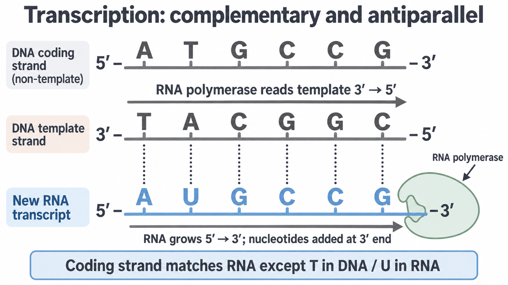
In the cell, step 5 means only the newest RNA stays paired with the template.
The coding strand is the non-template strand. Written in the same direction, it matches the RNA except for T in DNA and U in RNA. Do not replace T with U in the template sequence: complement it first.
From pre-mRNA to mature mRNA
In the AQA prokaryotic model, transcription produces mRNA directly: mRNA is its immediate product.
In eukaryotes, the immediate product is pre-mRNA, made in the nucleus and still containing the introns from 3.4.1. Introns do not code for amino acids, so if they stayed in, ribosomes would read them as codons and build the wrong polypeptide.
Splicing processes this RNA, removing introns and joining the exons, so exon 1–intron 1–exon 2 becomes exon 1–exon 2. The mature mRNA exits through a nuclear pore to reach cytoplasmic ribosomes. Splicing changes the RNA, not the chromosome's DNA.
When describing splicing, say that introns are removed and exons joined: “pre-mRNA is spliced” alone says too little.
Continue this lesson.
You’re previewing NovaLearn without an account. Create a free account to read the rest.
Create free account