3.8.1 - Mutations and protein structure
One changed base in the β-globin gene can cause sickle-cell disease, while another can leave the polypeptide untouched. Trace any gene mutation from DNA to polypeptide, and use a supplied genetic code to predict what it does.
From changed base to changed protein
A changed base can end up reshaping an enzyme's active site, but each link in that chain is only a possibility.
Gene mutation
A gene mutation is a change in the DNA base sequence of a gene.
Gene mutations might arise during DNA replication, when a copying error is not corrected. A changed amino acid sequence is a possible consequence, not part of the definition.
How a mutation can reach the protein
Follow a gene through to its protein's function:
- The gene's base sequence changes, so mRNA transcribed from it can carry a changed codon.
- Ribosomes read mRNA three bases at a time, so a changed codon may specify a different amino acid. This alters the primary structure, the sequence of amino acids.
- The new R group means hydrogen bonds, ionic bonds and disulfide bridges may form in different places.
- The polypeptide may therefore fold differently, changing its tertiary structure.
- An enzyme's active site may change shape, so the substrate is less complementary and binds less effectively.
Each "may" matters: a changed DNA sequence doesn't always change an amino acid, and a changed amino acid doesn't always stop a protein working. Other proteins follow the same chain, ending at a binding site or surface.
Keep the molecules separate
A code table normally gives mRNA codons read 5′ to 3′. If you are given DNA, first identify the strand and its direction.
| Molecule | Example | Link to mRNA |
|---|---|---|
| Template DNA | 3′–TAC CTT–5′ | Complementary to the mRNA |
| Coding DNA | 5′–ATG GAA–3′ | Same sequence as the mRNA, with T for U |
| mRNA | 5′–AUG GAA–3′ | Read by ribosomes |
DNA is transcribed and mRNA is translated. Bases code for amino acids; they do not turn into them.
Substitution
Swap one base and usually one codon changes, so the outcome depends on what that new codon specifies.
A substitution replaces one base with another. Each base belongs to one triplet and the number of bases stays the same, so within a coding sequence only one triplet changes.
Take mRNA codon GAA, which specifies glutamate. One substitution has three possible outcomes:
| New codon | Specifies | Effect on the polypeptide |
|---|---|---|
| GAG | Glutamate | None: a silent change, with the same amino acid sequence |
| GUA | Valine | One amino acid is replaced |
| UAA | Stop | Translation ends early, so the polypeptide is shorter |
The first row works because the genetic code is degenerate: several codons can specify the same amino acid. DNA and mRNA change, but the encoded amino acid is unchanged. Let the supplied code decide: don't assume that every mutation changes primary structure, an error AQA's 2024 examiners noted.
When one amino acid matters
In sickle-cell disease, a substitution changes β-globin mRNA GAG to GUG, replacing glutamate with valine. Glutamate's R group is charged and valine's is non-polar, so the surface of haemoglobin changes, and it can associate into fibres when deoxygenated. Where the change falls and the new R group's chemistry matter more than how many bases altered.
Addition, deletion and duplication
Lose one base and every later codon is regrouped; lose three and the rest of the gene stays in frame.
An addition inserts bases, a deletion removes bases, and a duplication repeats a DNA segment. To predict the effect, group the resulting mRNA from the same start point as before.
When the frame shifts
Ribosomes read codons with no gaps, in the reading frame set at the start codon (3.4.2). Add or remove one base, and every later base falls into a different position within its triplet.
Frame shift
A frame shift is a change to the reading frame, caused by adding or removing a number of bases that is not a multiple of three. It changes the nature of all base triplets downstream from the mutation.
Many amino acids may change, and a stop codon may appear at a different position. In the diagram, compare the rows: base 4 is missing from the lower one.
[DIAGRAM: asset_name: mutation-reading-frame-v2; asset_slug: mutation-reading-frame-v2; recommended_method: local_png; caption: Original and one-base-deleted mRNA with codon boundaries and a supplied genetic-code key.; description: Original and one-base-deleted mRNA with codon boundaries and a supplied genetic-code key.]
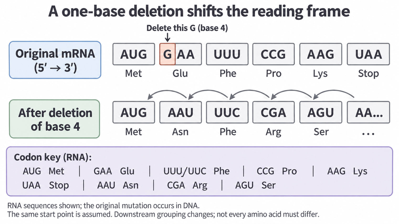
- After the deletion: every later base moves one place left. The mRNA now specifies Met–Asn–Phe–Arg–Ser and continues beyond the fragment, so its eventual length is unknown.
- The old stop: UAA is split between AGU and AA…, so it is no longer read as a codon.
- The third amino acid: it stays Phe, because UUU and UUC both specify it. All later triplets are regrouped, but not every amino acid changes.
When the frame is preserved
Adding or removing a multiple of three bases preserves the downstream frame. Whole triplets add or remove amino acids, and a change crossing a codon boundary can also alter the codon there. Duplication follows the same rule.
Inversion and translocation
Inversion and translocation rearrange whole stretches of DNA, so start by asking where the DNA broke.
Inversion
An inversion reverses the orientation of a DNA segment at its original location. The DNA breaks at two points, called breakpoints, and the segment rejoins the other way round, with its bases in reverse order.
Continue this lesson.
You’re previewing NovaLearn without an account. Create a free account to read the rest.
Create free account