3.2.1.1 - Eukaryotic cells
A pancreatic cell exports enzymes, a muscle cell contracts and a leaf cell captures light. Learn the structure and function of each organelle, then use them to explain specialised cells and how they build tissues, organs and systems.
The cell-surface membrane and nucleus
An oak tree, a mushroom and your own body are all built from the same basic kind of cell.
Cell theory is a unifying concept in biology: living organisms consist of cells, cells are the basic units of living organisation, and cells arise from existing cells.
These eukaryotic cells contain a nucleus and other organelles, structures with particular functions; many are membrane-bound compartments. Compartmentalisation allows different conditions and reactions to be maintained in different parts of the same cell.
How the membrane controls exchange
The cell-surface membrane surrounds the cytoplasm, inside any cell wall. It is a phospholipid bilayer (3.1.3) with embedded proteins, which explains its jobs:
- The hydrophobic centre slows water-soluble substances and ions, separating internal conditions from the external environment.
- Embedded proteins provide routes for particular substances, controlling what enters and leaves.
- Proteins at the surface allow cell recognition and cell signalling.
How the nucleus controls the cell
The nucleus is usually the most obvious organelle:
- The nuclear envelope is a double membrane around it.
- Nuclear pores in the envelope control exchange with the cytoplasm. mRNA and ribosomal subunits leave through them.
- Chromosomes consist of protein-bound, linear DNA. For most of the cell cycle it is spread out as chromatin; compact chromosome shapes are most visible during division.
- Nucleoplasm is the material surrounding the chromatin and nucleoli.
- One or more nucleoli make ribosomal RNA and assemble ribosomal subunits. A nucleolus is a region within the nucleus, not a separate membrane-bound organelle.
The nucleus acts as a control centre because genes in the DNA are used to make mRNA, which leaves through a pore and directs protein synthesis in the cytoplasm. The nucleus holds the instructions; ribosomes assemble the polypeptides.
Organelles for energy transfer
Two organelles handle a cell's main energy transfers: chloroplasts capture light and mitochondria make ATP.
Mitochondria and ATP
Mitochondria are the sites of the aerobic stages of respiration and of most ATP production. ATP then transfers energy to processes such as active transport and muscle contraction. Each mitochondrion has:
- a double membrane
- an inner membrane folded into cristae, which carries proteins involved in ATP synthesis
- a matrix containing enzymes for respiration, DNA and ribosomes
Folding gives a large surface area for these proteins, so more ATP can be made. Cells with high ATP demand, such as muscle cells and cells carrying out active transport, usually contain many mitochondria, often with densely packed cristae.
Watch out
Mitochondria do not create energy. Write that they produce ATP, because “produce energy” can lose marks.
Chloroplasts and light
Chloroplasts are the sites of photosynthesis, found in the photosynthetic cells of plants and algae. A chloroplast has:
- a double membrane envelope
- thylakoids, flattened membrane sacs containing chlorophyll, arranged in stacks; one stack is a granum and several are grana
- a stroma containing enzymes, DNA and ribosomes
The thylakoid membranes provide a large surface area for the light-dependent stage of photosynthesis. The stroma's enzymes carry out sugar production in the later stage.
Not all plant cells have chloroplasts. Palisade mesophyll cells near the upper leaf surface are packed with them, but most root cells have none: root hair cells absorb water and mineral ions underground, where little light reaches. Photosynthetic cells also respire, so they have mitochondria too.
| Mitochondrion | Chloroplast | |
|---|---|---|
| Process | Aerobic stages of respiration | Photosynthesis |
| Boundary | Double membrane | Double membrane envelope |
| Internal membranes | Cristae: folds of the inner membrane | Thylakoids: stacked into grana |
| Fluid and its enzymes | Matrix: respiration | Stroma: sugar production |
| Also contains | DNA and ribosomes | DNA and ribosomes |
Cover the table, then compare the two organelles from memory.
Making, modifying and moving substances
Follow one digestive enzyme out of a pancreatic cell and you meet the organelles of secretion in order.
Ribosomes and the endoplasmic reticulum
Ribosomes are the site of protein synthesis: small particles of ribosomal RNA and protein, with one large and one small subunit and no membrane. Those free in the cytoplasm or attached to the rough endoplasmic reticulum (RER) are 80S; smaller 70S ribosomes occur inside mitochondria and chloroplasts.
Free ribosomes make many proteins used within the cytoplasm; RER-bound ribosomes make many exported or membrane proteins, such as digestive enzymes.
The rough endoplasmic reticulum (RER) is a network of flattened sacs called cisternae, with ribosomes attached to its outer surface. It provides a large surface area for protein synthesis, folds and transports the proteins made there, and is especially abundant in secretory cells.
The smooth endoplasmic reticulum (SER) is a network of membrane tubules with no ribosomes. It synthesises, stores and transports lipids and also handles some carbohydrate processing.
The Golgi apparatus and secretion
The Golgi apparatus is a more compact stack of flattened membranes. It receives proteins and lipids from the ER, then:
- modifies them, for example by adding carbohydrate to a protein to form a glycoprotein
- labels and sorts them
- packages them into Golgi vesicles, small membrane-bound sacs that bud off the stack
- forms lysosomes
Find the rough ER in the diagram, then follow the arrows to the secreted protein on the right.
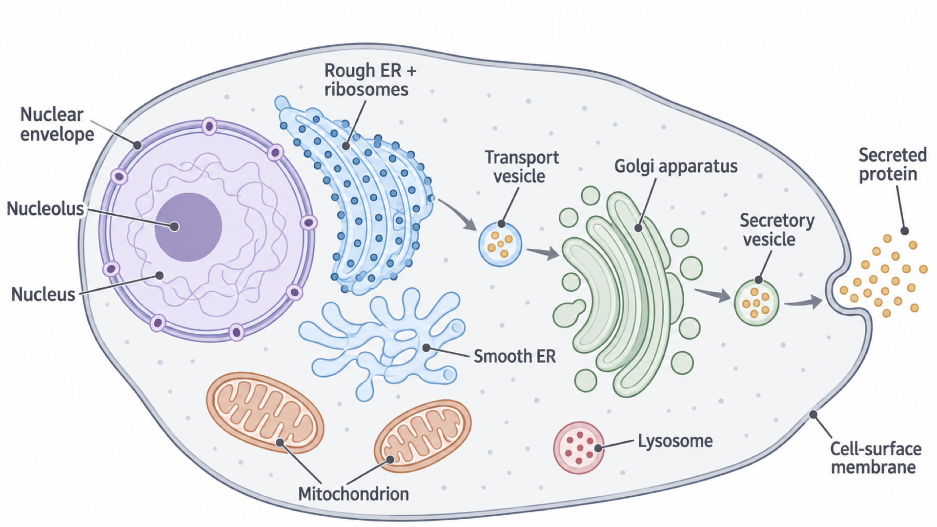
- A ribosome on the RER makes a polypeptide, using mRNA from the nucleus.
- The polypeptide enters the RER, which folds and transports it.
- Transport vesicles carry it from the RER to the Golgi apparatus.
- The Golgi apparatus modifies, sorts and packages it.
- A secretory vesicle (a Golgi vesicle bound for the cell surface) moves to the cell-surface membrane.
- Vesicle and membrane are both phospholipid bilayers, so they fuse, releasing the enzyme by exocytosis.
Exam tip
In a question on the role of organelles in making and releasing an enzyme, the nucleus (DNA codes for it) and mitochondria (they produce ATP) can also earn marks.
Lysosomes and controlled digestion
Lysosomes are single-membrane organelles formed by the Golgi apparatus. Their hydrolytic enzymes can digest worn-out organelles, unwanted cell components and material engulfed by phagocytosis.
A lysosome can fuse with a vesicle containing engulfed material and release its enzymes into that compartment. Membranes keep the enzymes from the rest of the cytoplasm, so digestion stays controlled.
Cell walls and the plant vacuole
In pure water a red blood cell bursts, but a plant cell simply becomes firm.
What cell walls are made of
A cell wall lies outside the cell-surface membrane in plants, algae and fungi. Animal cells lack one. Wall composition varies between groups:
- plant cell walls contain cellulose (3.1.2)
- many algal walls contain cellulose; others contain different materials including glycoproteins
- fungal cell walls contain chitin, glycan and glycoproteins
Cell walls:
- provide mechanical strength, supporting the organism as a whole
- help prevent osmotic lysis, the bursting of a cell when water enters by osmosis
- in plants, allow water to move along cell walls
How the vacuole makes a cell firm
A mature plant cell usually has one large central vacuole surrounded by a membrane, the tonoplast. The vacuole contains cell sap, a solution of mineral salts, sugars, amino acids, wastes and sometimes pigments. It stores solutes, its pigments can contribute to petal colour, and it keeps the cell turgid:
- In dilute surroundings, the solutes inside keep the cell's water potential lower than outside, so water enters by osmosis.
- The vacuole expands and the contents press against the wall.
- The wall resists further expansion, which stops the cell bursting and produces turgor pressure.
- Turgor pressure supports the cell.
A red blood cell has no wall to resist expansion, so it can burst. If a plant cell loses water, turgor pressure falls and the tissue may become flaccid.
In the diagram, work inwards from the wall to the cell-surface membrane, then the tonoplast.
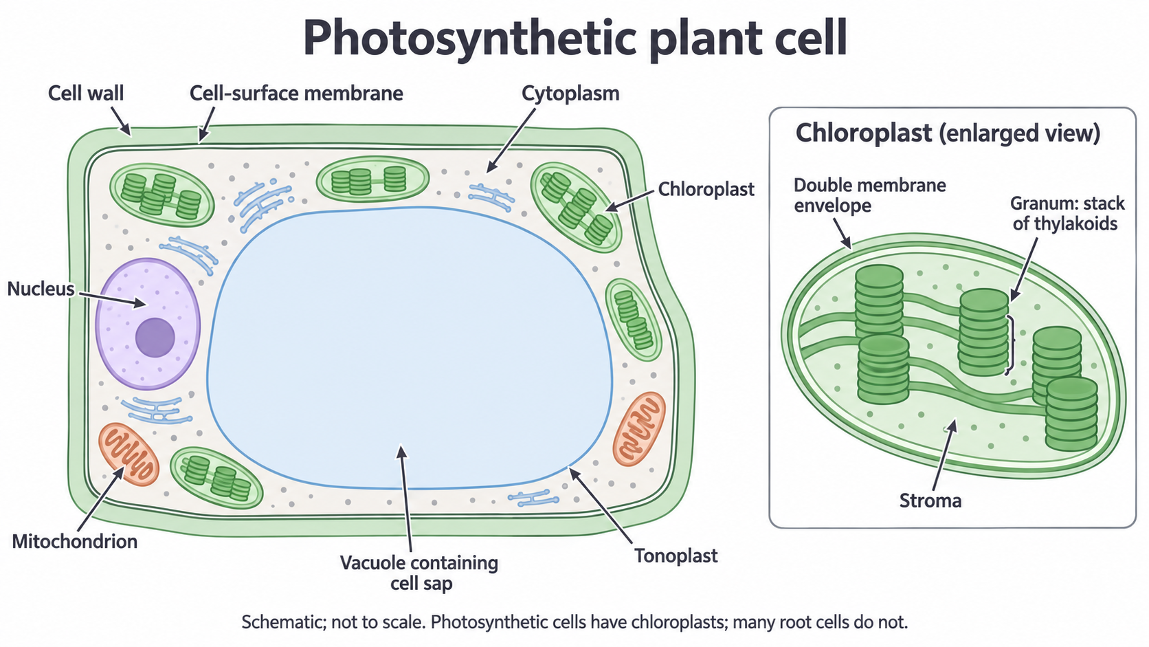
The wall does not replace the membrane:
| Boundary | Surrounds | Role |
|---|---|---|
| Cell wall | The cell-surface membrane | Strength; freely permeable to many substances |
| Cell-surface membrane | The cytoplasm | Selective: controls what enters and leaves the cell |
| Tonoplast | The vacuole | Separates cell sap from the cytoplasm |
From specialised cells to whole organisms
A muscle cell and a pancreatic cell carry the same genes but do very different jobs.
Why specialised cells differ
In complex multicellular organisms, eukaryotic cells become specialised. Most body (somatic) cells contain the same genes (a mature mammalian red blood cell, which has lost its nucleus, is an exception), but different genes are expressed in different cell types. This gives rise to different structures and combinations of organelles.
So a cell's organelles reflect the work it does:
- palisade mesophyll cells contain many chloroplasts, which absorb light for photosynthesis
- muscle cells contain many mitochondria, which produce ATP for contraction
- phagocytes contain many lysosomes, whose hydrolytic enzymes digest engulfed pathogens
- secretory cells have extensive RER and a large Golgi apparatus to make and export proteins, and many mitochondria because protein synthesis and vesicle movement use ATP
“Many mitochondria because the cell is active” is incomplete: state which ATP-requiring process is supported.
Organelle abundance can suggest a function, but usually does not identify a unique cell type by itself. Pancreatic cells and antibody-secreting plasma cells both have extensive RER, for example.
From tissues to organ systems
Specialised cells are organised into tissues, tissues into organs and organs into systems:
- a tissue is an organised group of cells working together for a particular function; it may include more than one cell type
- an organ is a group of tissues working together
- an organ system is a group of organs working together
Palisade mesophyll, spongy mesophyll, xylem and phloem are tissues that together form a leaf, an organ. In animals, tissues form organs such as the stomach or heart, and organs form systems such as the digestive or circulatory system.