3.6.1.3 - Control of heart rate
This lesson follows the wave of electrical excitation that starts each heartbeat, then shows how receptors and the autonomic nervous system change how often it happens, and how to measure the result.
Myogenic stimulation of the heart
Each heartbeat starts inside the heart, and a fixed route makes the atria contract before the ventricles.
A transplanted heart has had its nerves from the brain cut, yet it beats in its new chest. So the stimulus for each beat must come from the heart itself.
Myogenic
Describes muscle that initiates its own contraction without external nervous stimulation.
Cardiac muscle is myogenic, so the basic rhythm starts within the heart rather than being triggered by motor neurones. The SAN, AVN and Purkyne tissue below are specialised cardiac muscle, not neurones.
The route of excitation
Start at the top left of the diagram, the heart's right side. Orange arrows show excitation, the yellow band is non-conductive tissue, and blue lines are the bundle of His and Purkyne tissue.
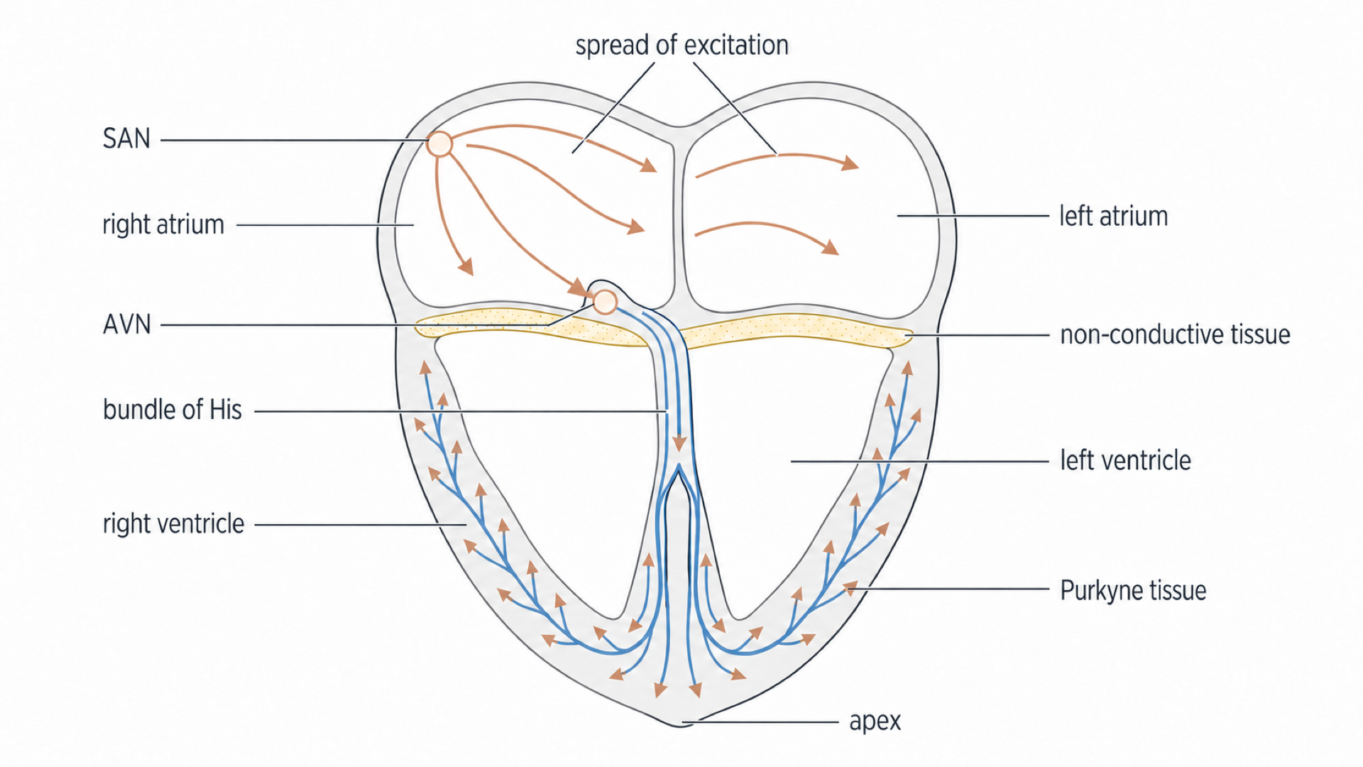
- The sinoatrial node (SAN) in the wall of the right atrium initiates a wave of electrical excitation. Because it sets the basic rhythm, it is the heart's pacemaker.
- The wave spreads across both atria, so the atria contract.
- A layer of non-conductive tissue between the atria and ventricles prevents the wave passing straight into the ventricles.
- The excitation reaches the atrioventricular node (AVN), which delays it briefly. During the delay the atria finish contracting and the ventricles finish filling with blood.
- The AVN passes the excitation down the bundle of His, which is made of specialised conducting fibres called Purkyne tissue, to the apex of the heart, its pointed lower tip.
- Branches of Purkyne tissue carry the excitation up through the ventricular walls, so the ventricles contract together from the apex upwards. The aorta and pulmonary artery leave from the top of the ventricles, so this forces blood out of the heart.
Exam tip
In a 5-mark question on this sequence in 2024, a significant minority left out atrial contraction, and some left out the AVN delay. “Wave of excitation”, “electrical activity”, “depolarisation” and “impulses” were accepted, but “signal” or “message” can lose a mark.
Why the order matters
The non-conductive tissue and the AVN delay coordinate the two contractions. If the atria and ventricles contracted together, the ventricles would fill less completely and less blood would be pumped out in each beat.
The autonomic nervous system and the SAN
The SAN sets the rhythm, but two opposing sets of nerves from the brain change how fast it runs.
Heart rate changes between rest and exercise because the medulla oblongata in the brain alters it through the autonomic nervous system.
Autonomic nervous system
The part of the nervous system that controls involuntary activities of internal muscles and glands. It has sympathetic and parasympathetic divisions.
The two branches are antagonistic on heart rate, and each is driven by its own centre in the medulla oblongata:
- Sympathetic nervous system: the acceleratory centre increases the frequency of impulses to the SAN along the sympathetic nerve, which increases heart rate.
- Parasympathetic nervous system: the inhibitory centre increases the frequency of impulses to the SAN along the vagus nerve, which decreases heart rate.
The SAN as effector
In this control system, the effector is the SAN, the tissue these nerves act on. The impulses do not start each heartbeat. They alter the frequency at which the SAN generates waves of electrical excitation, and each wave triggers one heartbeat, so this frequency sets the heart rate.
Both branches are active even at rest, so heart rate depends on the balance between them. At rest the parasympathetic side dominates, which holds the SAN below its own rate.
A transplanted heart shows this: surgery cuts its vagus connection, so its resting rate is raised, commonly to about 90 to 110 beats per minute.
Blocking one branch
A drug that blocks parasympathetic action at the SAN removes an inhibitory influence, so the SAN generates waves more frequently without any extra sympathetic stimulation. When AQA asked this about the drug atropine in 2025, relatively few answers named the SAN, which was a mark.
Chemoreceptors and blood pH
Hard exercise lowers blood pH, and receptors in your arteries respond by raising heart rate.
Chemoreceptors
Receptors in the walls of the carotid arteries and the aorta that detect changes in blood pH associated with changes in carbon dioxide concentration.
As in the Bohr effect, carbon dioxide forms carbonic acid in blood, increasing hydrogen-ion concentration and lowering pH. In hard exercise, lactate from anaerobic respiration lowers pH too. Chemoreceptors detect this chemical change. They are distinct from the stretch-sensitive pressure receptors in the same regions.
From low pH to a faster heart
- During exercise, respiration increases and more carbon dioxide is produced.
- More carbon dioxide in the blood lowers blood pH.
- Chemoreceptors in the carotid arteries and aorta detect the fall in pH.
- They send impulses at a higher frequency to the acceleratory centre in the medulla oblongata.
- The medulla oblongata sends impulses at a higher frequency along the sympathetic pathway to the SAN.
- The SAN, acting as the effector, generates waves of excitation more frequently, so heart rate rises.
- Faster circulation delivers carbon dioxide to the lungs, where ventilation removes it, so blood pH returns towards normal.
Watch out
More carbon dioxide lowers blood pH. On a 2025 paper some students wrote that it raises pH.
Negative feedback
This is negative feedback: the response opposes the original change. As pH returns towards its resting value, chemoreceptors send fewer impulses, so sympathetic impulses to the SAN become less frequent and heart rate falls back towards its resting rate.
The fall in pH also stimulates chemoreceptors to increase ventilation rate, so a question about breathing needs that route, not heart rate.
Pressure receptors and blood pressure
Higher blood pressure stretches artery walls further, and receptors there turn that stretch into impulses.
Pressure receptors
Receptors in the walls of the carotid arteries and the aorta that detect changes in blood pressure.
They sit in the carotid sinuses (widened parts of the carotid arteries) and the aortic arch. As with the Pacinian corpuscle in 3.6.1.2, a stronger stimulus gives a higher impulse frequency, so more stretch sends more impulses to the medulla oblongata.
Continue this lesson.
You’re previewing NovaLearn without an account. Create a free account to read the rest.
Create free account