3.8.2.3 - Gene expression and cancer
Tumours develop when the controls on cell division fail. Learn how faulty genes, methylation and oestrogen cause this, and how to judge evidence about risk and cure.
Benign and malignant tumours
Tumours form when cells divide out of control, but only malignant ones invade and spread.
A tumour is an abnormal mass of cells, formed when cell production outstrips cell loss. Uncontrolled cell division by mitosis is a key feature.
Benign tumours stay localised: they do not invade neighbouring tissue or spread to distant sites. They can still be dangerous, by pressing on vital structures or producing abnormal amounts of a hormone.
Malignant tumours are cancers. They invade surrounding tissue and can spread, because changes in cell adhesion and movement let cells detach.
How a malignant tumour spreads
Metastasis is the spread of cancer cells to form secondary tumours in distant tissue. Follow stages 1 to 4 on the right of the diagram.
[DIAGRAM: asset_name: tumour-invasion-v2; asset_slug: tumour-invasion-v2; recommended_method: local_png; caption: Malignant cells invade tissue, enter circulation and leave a vessel before growing as a secondary tumour at a distant site.; description: Malignant cells invade tissue, enter circulation and leave a vessel before growing as a secondary tumour at a distant site.]
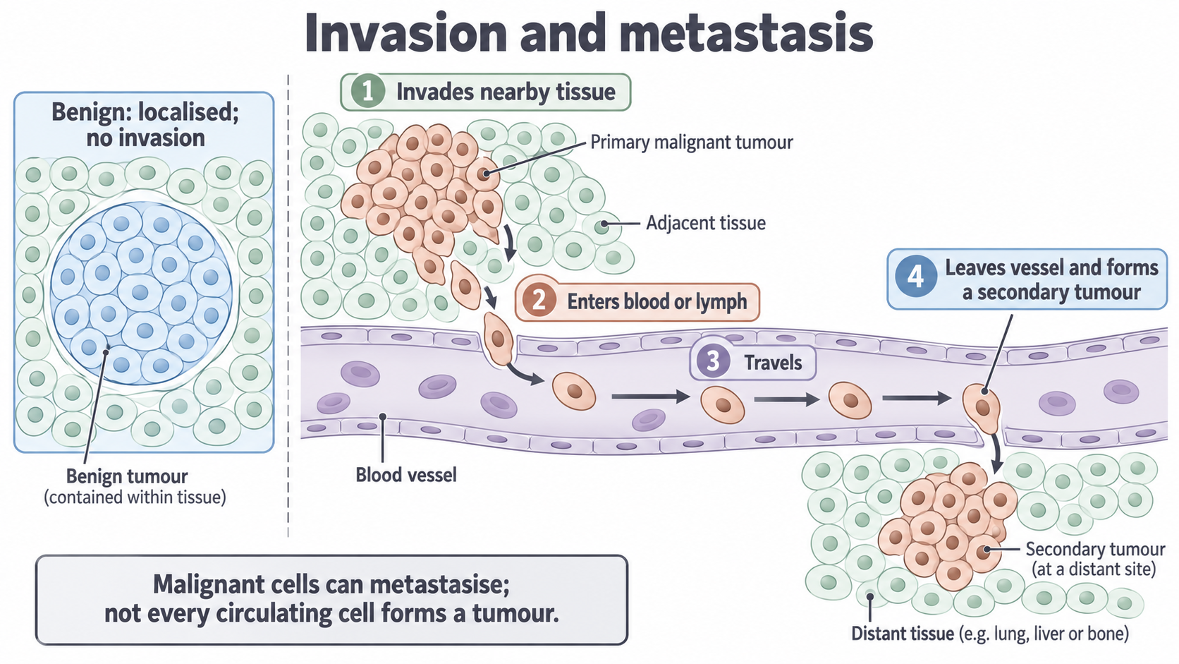
- Cells leave the primary tumour and invade nearby tissue.
- They enter the blood or lymph.
- They travel in the circulation.
- They leave the vessel and grow into a secondary tumour in distant tissue.
Entering the circulation is not enough, because many cells fail to survive or grow at the new site.
Invasion and metastasis are the central distinction; the other rows of this table are usual patterns, not absolute rules. Pair each feature when you compare: benign tumours do not invade, whereas malignant tumours do.
| Feature | Benign tumour | Malignant tumour |
|---|---|---|
| Invasion and metastasis | Neither | Invades; can metastasise |
| Growth rate | Usually slow | Usually faster |
| Cells and nuclei | Usually well differentiated (specialised), with normal nuclei | Often poorly differentiated, with larger, irregular nuclei |
| Adhesion | Adhesion molecules hold cells together | Often fewer adhesion molecules, so cells break away |
| Capsule | Some have one | Usually none |
| After removal | Rarely returns | More likely to return |
Cover the table and recall its rows.
Oncogenes and proto-oncogenes
Normal genes that drive cell division can become oncogenes, which keep the signal to divide switched on.
Division normally starts when a growth factor binds a receptor in the cell-surface membrane, and signalling proteins inside the cell then stimulate the cell cycle. Proto-oncogenes code for proteins such as growth factors, receptors and signalling proteins.
Proto-oncogene and oncogene
A proto-oncogene is a normal gene whose product stimulates regulated cell growth or division. An oncogene is a mutated or overexpressed form that stimulates excessive growth or division.
Two routes to too much signal
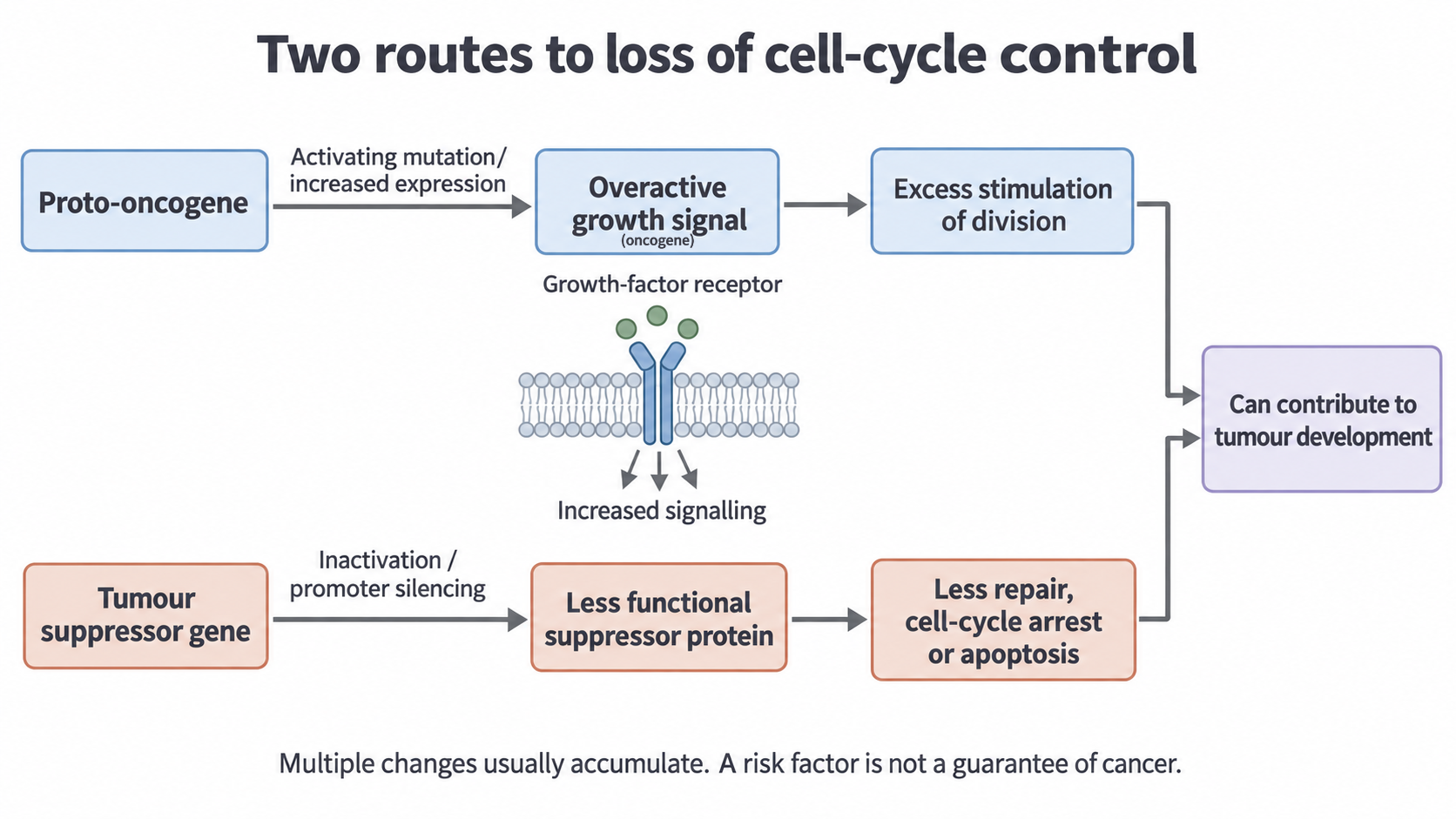
- Activating mutation. A mutated receptor, for example, can signal continuously, even without growth factor.
- Excessive gene expression. The cell makes too much growth-promoting protein, such as a growth factor or receptor, for example because it has extra copies of the gene, so more mRNA is transcribed.
Either route overstimulates the cell cycle, so cells divide too often.
This is a gain of function: the altered or overproduced product actively promotes signalling, so one altered allele can sometimes be enough, even beside a normal allele. That does not make cancer certain, because tumours commonly involve several accumulating changes.
The oncogene is DNA, while the receptor or growth factor is protein, so a drug can block the product's activity without changing the DNA sequence.
Tumour suppressor genes
Tumour suppressor genes restrain damaged cells, so losing one can let those cells survive and keep dividing.
A tumour suppressor gene encodes a product that helps prevent uncontrolled growth. A mutation can stop it producing working protein. This loss of function removes a control on damaged cells. Products differ, and one need not perform all three roles:
- Cell-cycle arrest pauses the cycle. Without it, a damaged cell divides.
- DNA repair corrects damage. Without it, more mutations persist.
- Apoptosis, programmed cell death, removes dangerous cells. Without it, they survive.
TP53 acts in damage responses, and BRCA1 and BRCA2 contribute to DNA repair.
Why one faulty allele raises risk
For many tumour suppressor genes, one working allele supplies enough function, so both copies must be inactivated for the full effect.
A person inheriting one faulty allele has it in every cell, so one further event in any cell can remove the function. Without that inheritance, both alleles must be inactivated in the same cell, which is rarer. Inheriting one faulty allele therefore raises risk, and tumours tend to appear earlier and in more places.
This is a common model, not a rule for every gene, and it does not make cancer certain.
Two routes compared
The diagram sets the routes side by side: gain of a signal on top, loss of a restraint below. Promoter silencing comes in the next section.
[DIAGRAM: asset_name: cancer-gene-control-v2; asset_slug: cancer-gene-control-v2; recommended_method: local_png; caption: Excess growth-promoting signalling and loss of tumour-suppressor function are distinct routes that can contribute to tumour development.; description: Excess growth-promoting signalling and loss of tumour-suppressor function are distinct routes that can contribute to tumour development.]
| Feature | Oncogene | Faulty tumour suppressor gene |
|---|---|---|
| Normal gene's role | Stimulates division | Restrains growth or repairs DNA |
| Change | Gain of function | Loss of function |
| Protein | Overactive or in excess | Reduced, absent or not working |
| Altered alleles for an effect | One can be enough | Often both |
Abnormal methylation and cancer
A gene's base sequence can stay unchanged while abnormal DNA methylation alters how much of it is expressed.
In 3.8.2.2 you saw that added methyl groups can reduce transcription. Hypermethylation means increased DNA methylation; hypomethylation means decreased DNA methylation.
Continue this lesson.
You’re previewing NovaLearn without an account. Create a free account to read the rest.
Create free account