3.6.2.1 - Nerve impulses
A nerve impulse arrives at full size because the axon rebuilds it on the way. Trace how ion gradients produce it, why myelin speeds it up, and how the refractory period limits its frequency.
Structure of a myelinated motor neurone
One motor neurone can run from the lower spinal cord to the big toe, some 1.3 m, yet it is a single cell.
A motor neurone carries nerve impulses from the central nervous system to an effector, such as a skeletal muscle. Follow the upper drawing from left to right, the direction the impulse travels.
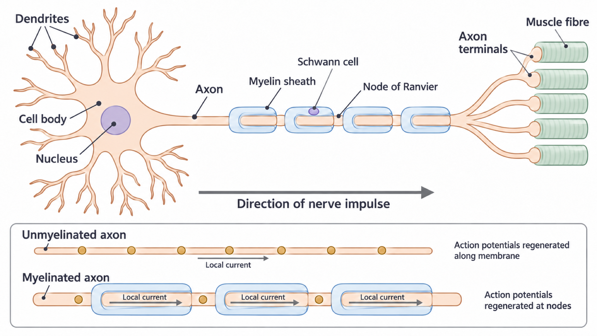
- Branched dendrites receive input from other neurones.
- The cell body contains the nucleus and the organelles that maintain the cell.
- A single axon carries action potentials away from the cell body.
- Branched axon terminals end at muscle fibres, where neurotransmitter can be released.
Schwann cells, myelin and nodes
In a peripheral nerve, each Schwann cell wraps its membrane repeatedly around a short length of axon. The layers form the insulating myelin sheath, so each blue segment is one Schwann cell. The Schwann cell is the whole supporting cell, with its own nucleus (the purple oval); myelin is the layered membrane it produces.
Small gaps between successive myelin segments are nodes of Ranvier. They contain many voltage-gated ion channels (channels that open or close as membrane voltage changes), so action potentials can be regenerated there.
The sheath greatly reduces ion movement across the covered membrane, but it does not block current flowing along the inside of the axon.
In the lower strips, dots mark successive sites of regeneration, not separate cells, and local current flows inside the continuous axon, not through the myelin.
Exam tip
On a labelled neurone, “node” alone can lose a mark: write “node of Ranvier”. AQA did not credit “myelin sheath” for a Schwann cell in 2025.
The resting potential
A resting neurone is not idle: it spends ATP constantly, maintaining the ion gradients behind its negative interior.
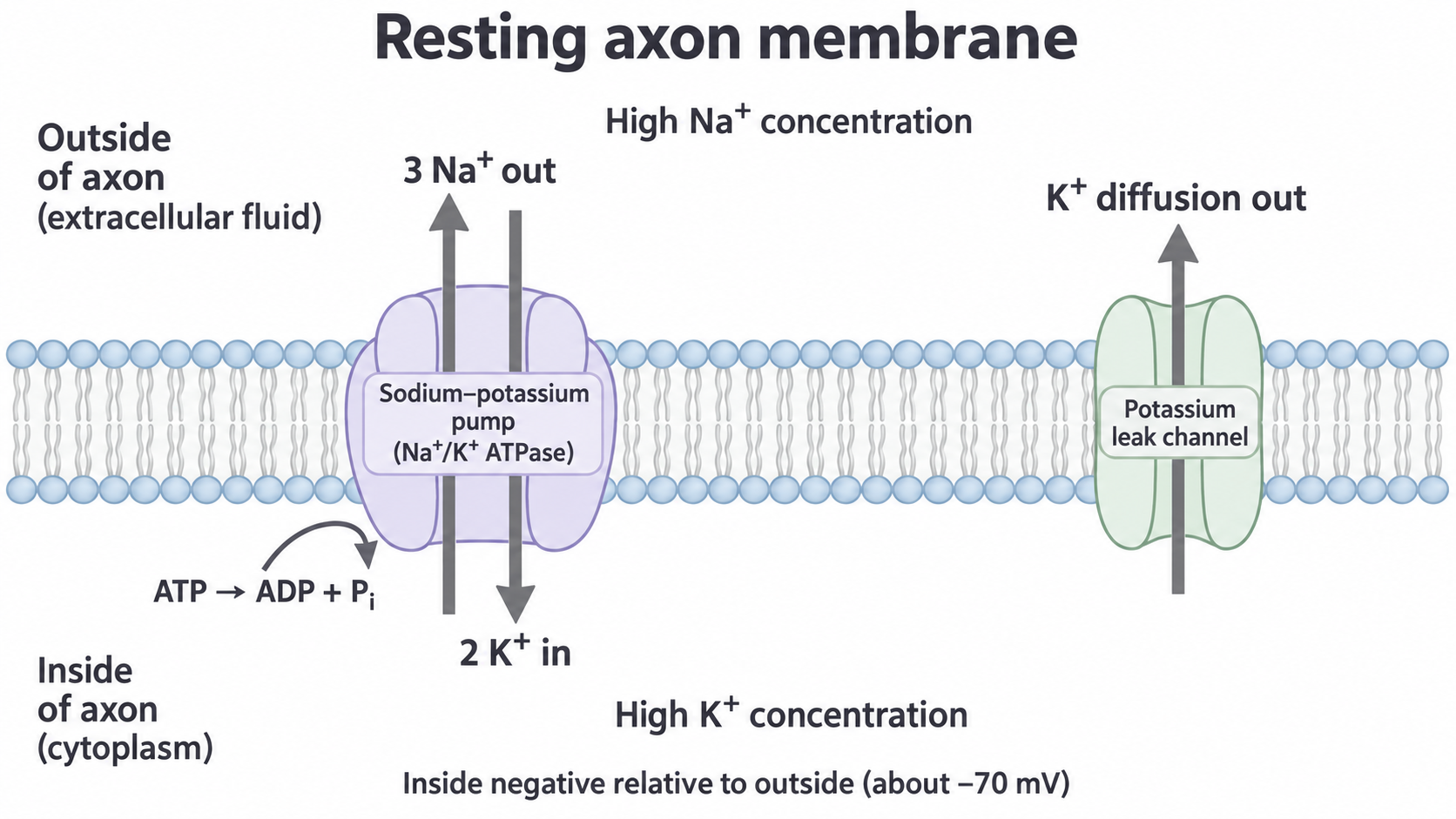
The inside is negative relative to the outside, so the membrane is electrically polarised. Membrane potential is this potential difference, and the resting potential is often about −70 mV. It describes a difference across the membrane, not a negative charge on the entire cell.
Gradients maintained by the pump
The sodium–potassium pump hydrolyses ATP to transport three sodium ions out for every two potassium ions in. They move against their concentration gradients, so this is active transport. Sodium-ion concentration stays high outside and potassium-ion concentration high inside. The unequal exchange adds slightly to the negative interior.
If the pump is inhibited, sodium ions keep diffusing in and potassium ions keep diffusing out through channels. The gradients are no longer maintained, so over time the ion concentrations on each side become more similar and the resting potential is lost.
Selective permeability and electrical attraction
The membrane is much more permeable to potassium than sodium. This differential membrane permeability produces most of the charge difference:
- Potassium leak channels are open, whereas most voltage-gated sodium channels are closed.
- Potassium ions diffuse out down their concentration gradient, leaving a small excess of negative charge inside near the membrane.
- The negative inside attracts potassium ions back, increasingly opposing further loss.
An electrochemical gradient combines the concentration difference and the electrical attraction or repulsion. For potassium they oppose each other: concentration pushes ions out while charge attracts them back in. For sodium both point inwards, but low permeability limits entry.
Resting potential is a stable balance of ion movements, not a state in which all ion movement stops. In the drawing, potassium crosses both ways: in through the pump, out through the leak channel.
| Ion | Moved by the pump | Channels at rest | Result |
|---|---|---|---|
| Sodium | 3 out | Mostly closed | High outside, little enters |
| Potassium | 2 in | Leak channels open | High inside, diffuses out, leaves inside negative |
Cover the table, then explain the resting potential from memory.
Depolarisation and the action potential
Each phase of an action potential traces back to a change in membrane permeability.
An action potential is a brief, rapid change in membrane potential that can be regenerated along an axon. The graph follows one membrane region through time, not different places along the axon.
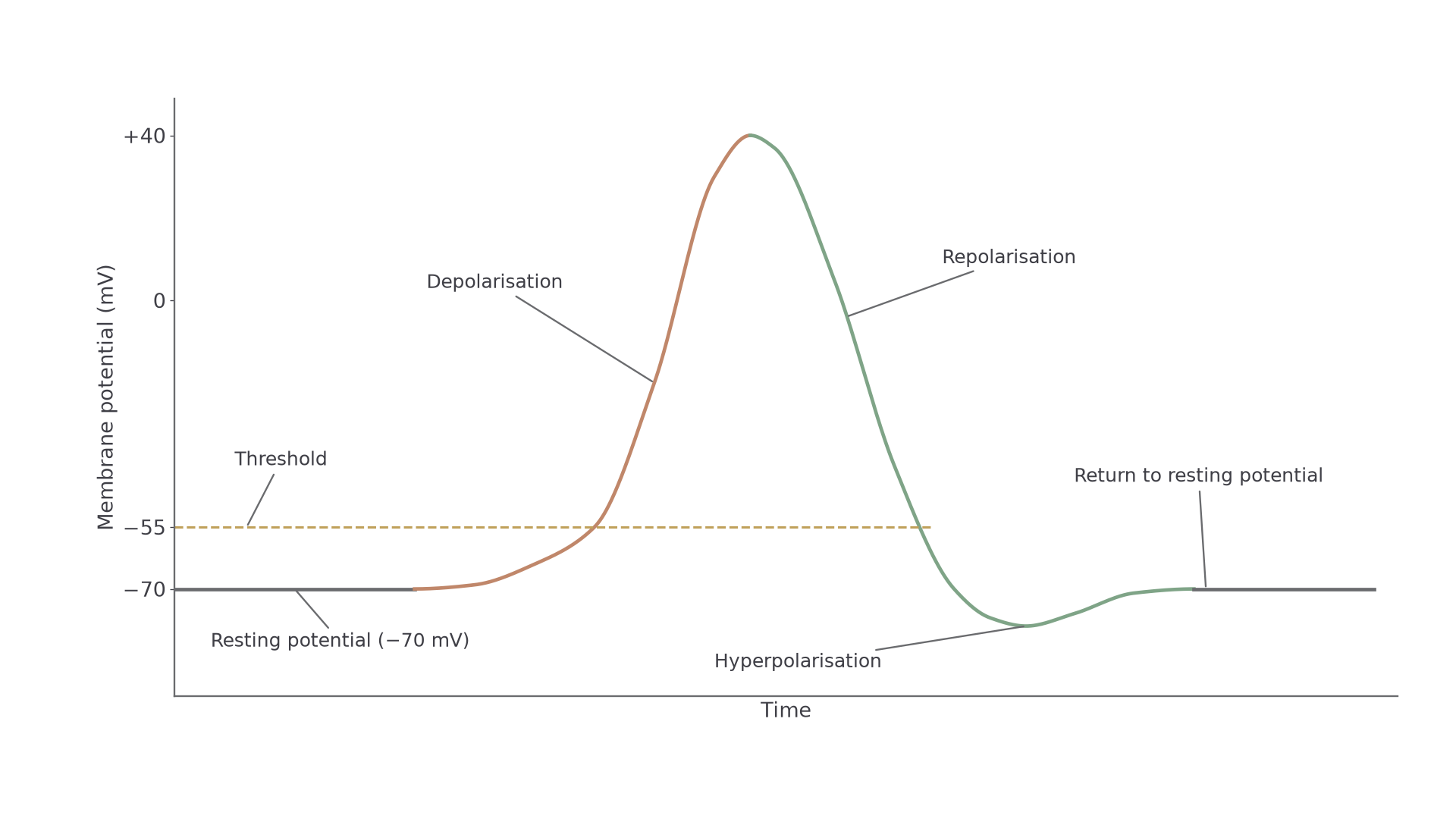
Read the order of events, not a duration: the time axis has no scale, and the values are typical. The brown rise is sodium entering; the green fall is potassium leaving.
The rise: sodium permeability increases
- A receptor’s generator potential (3.6.1.2) or an excitatory input at a synapse (next lesson) makes the inside less negative: a local depolarisation.
- If this brings a trigger region to threshold (−55 mV on this graph), voltage-gated sodium channels open rapidly, so sodium permeability rises.
- Sodium ions enter by facilitated diffusion down their electrochemical gradient.
- Positive charge entering causes further depolarisation, opening more sodium channels. This positive feedback produces the steep rise, and the inside briefly becomes positive, often roughly +30 to +40 mV.
The fall: potassium permeability increases
- Voltage-gated sodium channels become inactivated: closed, and unable to reopen until the membrane recovers. Sodium entry largely stops.
- Voltage-gated potassium channels open, so potassium ions leave, making the interior negative again. This is repolarisation.
- These channels close with a delay, so potassium efflux continues and the membrane becomes more negative than at rest. This is hyperpolarisation.
- As potassium permeability returns to its resting level, so does the potential, and sodium channels recover.
Watch out
The sodium–potassium pump does not cause the fast fall. It continuously maintains the gradients, and one action potential moves too few ions to reverse them.
Both phases are diffusion through channels, so neither uses ATP directly.
Continue this lesson.
You’re previewing NovaLearn without an account. Create a free account to read the rest.
Create free account