3.6.4.2 - Control of blood glucose
Blood glucose rises after a meal and falls during a fast, yet it stays within narrow limits. Learn how the liver and three hormones hold it there, what fails in diabetes, and how to judge glucose data and claims about Type II diabetes.
Why blood glucose concentration changes
Blood glucose behaves like a balance: it rises when glucose enters the plasma faster than it leaves.
Blood glucose concentration
The concentration of glucose in the blood plasma. In humans it is kept close to about , although it fluctuates around this value.
These are the main flows in and out of the plasma:
| Glucose enters the plasma | Glucose leaves the plasma |
|---|---|
| Digestion of carbohydrate and absorption of glucose from the small intestine | Taken up by body cells for respiration |
| Released by the liver from glycogen (glycogenolysis) | Stored as glycogen in liver and muscle cells (glycogenesis) |
| Made by the liver from non-carbohydrates (gluconeogenesis) | Converted to lipids when glucose is in excess |
Four everyday factors change blood glucose concentration by altering these flows:
- Meal composition: a carbohydrate-rich meal adds more glucose to the blood than a meal of mostly protein or fat.
- Fasting: no glucose is absorbed from the gut, so the liver becomes the main supply while cells keep using glucose.
- Physical activity: exercise speeds up respiration in muscle, so glucose is used faster.
- Stress: adrenaline can raise blood glucose by triggering glycogen breakdown.
Watch out
Liver glycogen can help maintain blood glucose concentration, but muscle glycogen does not directly release glucose into the blood. Muscle cells break it down to glucose phosphate but lack the enzyme that removes the phosphate, so it stays in the cell as a local store for respiration.
The liver and the action of insulin
After a meal, insulin and the liver move glucose out of the blood and into storage.
Three liver processes
Glycogen is a large, insoluble storage molecule, so it has far less osmotic effect than an equivalent store of many separate glucose molecules. Hepatocytes (liver cells) can therefore store glucose after feeding and release it between meals, using three processes.
| Process | Conversion | Word clue |
|---|---|---|
| Glycogenesis | Glucose → glycogen | -genesis: making |
| Glycogenolysis | Glycogen → glucose | -lysis: splitting |
| Gluconeogenesis | Non-carbohydrates such as glycerol and amino acids → glucose | neo-: new |
Exam tip
Weaker answers in AQA's 2024 Paper 2 wrote "glycolysis", "glycogenesis" or "glucogenesis" for glycogen breakdown. "Glycogenolysis" or "glycogen hydrolysed to glucose" earned the mark.
How insulin lowers blood glucose
When blood glucose concentration rises, beta cells in the islets of Langerhans in the pancreas detect the change and secrete insulin. Insulin travels in the blood to target cells such as liver, muscle and adipose (fat-storing) cells. A target cell has a receptor complementary in shape to the hormone, so cells without that receptor do not respond.
Insulin first attaches to receptors on the surfaces of target cells. Two effects follow:
- More glucose enters muscle and adipose cells. Binding causes vesicles containing glucose transport proteins to fuse with the cell-surface membrane. More glucose then enters by facilitated diffusion, down its concentration gradient. The specification calls these channel proteins; strictly they are carriers, commonly called GLUT4.
- More glucose is stored in liver and muscle cells. Insulin activates enzymes involved in converting glucose to glycogen (glycogenesis).
Liver cells differ: their glucose transport is not controlled by insertion of GLUT4. Turning glucose into glycogen keeps the free glucose concentration inside the cell low, so glucose keeps diffusing in from the blood.
Overall, more glucose leaves the blood, and some is respired faster or converted to fat. As blood glucose concentration falls back towards the norm, the stimulus to the beta cells shrinks, so they secrete less insulin. This is negative feedback.
Glucagon and adrenaline
When glucose falls or a threat appears, glucagon and adrenaline raise it, but their triggers differ.
Glucagon corrects a fall
When blood glucose concentration falls, pancreatic alpha cells detect the change and secrete glucagon. Glucagon attaches to receptors on target cells, especially hepatocytes, and raises blood glucose concentration by activating enzymes involved in two conversions:
- glycogen to glucose (glycogenolysis)
- glycerol and amino acids into glucose (gluconeogenesis)
Watch out
Glucagon and glycogen are easy to swap. Don't write "glucagon is stored in the liver" or "the pancreas releases glycogen". Glucagon is the hormone from alpha cells; glycogen is the storage polysaccharide in liver and muscle cells. Also, glucagon doesn't convert glycogen to glucose itself. Write that it activates the enzymes that do.
Insulin and glucagon act antagonistically: insulin lowers blood glucose concentration and glucagon raises it. As glucose recovers, the stimulus for glucagon secretion shrinks, so alpha cells secrete less; falling glucose reduces insulin secretion in the same way. Each corrective response diminishes as glucose returns towards the norm.
Adrenaline responds to stress
Adrenaline is released from the adrenal glands in situations such as stress, fear or exercise. Its main trigger is the situation, not a low glucose reading, so don't treat it as part of the insulin–glucagon loop.
Adrenaline attaches to receptors on target cells and activates enzymes involved in converting glycogen to glucose. In the liver, this rapidly adds glucose to the blood, making more available for respiration during the fight-or-flight response. In muscle, the glycogen fuels local respiration instead.
| Insulin | Glucagon | Adrenaline | |
|---|---|---|---|
| Source | Beta cells | Alpha cells | Adrenal glands |
| Trigger | Glucose rises | Glucose falls | Stress, fear or exercise |
| Main effect | More glucose uptake; glycogenesis | Glycogenolysis; gluconeogenesis | Glycogenolysis |
| Blood glucose | Falls | Rises | Rises |
Cover the table, then give each hormone's source, trigger and effect from memory.
The second messenger model
Glucagon and adrenaline stay outside the cell, so how do they switch on enzymes inside it?
They are water-soluble hormones, so they do not pass through the phospholipid bilayer of the cell-surface membrane.
Second messenger
An intracellular signalling molecule produced after a hormone binds to a receptor on the cell-surface membrane. In this topic, cAMP is the second messenger.
The hormone is the first messenger because it carries the signal from elsewhere in the body; cAMP carries it inside the cell.
The pathway in a liver cell
Start at glucagon on the left of the diagram and follow the arrows to the glucose released at the bottom right.
[DIAGRAM: asset_name: Glucagon signalling - rebuilt; asset_slug: Glucagon signalling - rebuilt; recommended_method: retained_png; caption: Glucagon triggers adenylate cyclase, cAMP and protein kinase inside a liver cell, increasing glycogen breakdown.; description: Glucagon triggers adenylate cyclase, cAMP and protein kinase inside a liver cell, increasing glycogen breakdown.]
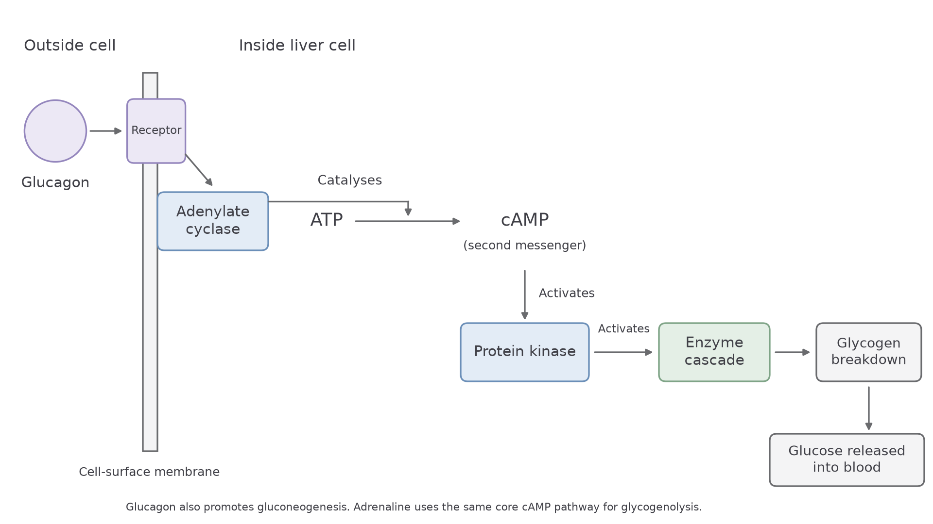
- The hormone binds to a specific receptor on the cell-surface membrane.
- This activates the membrane enzyme adenylate cyclase.
- Adenylate cyclase catalyses the conversion of ATP to cyclic AMP (cAMP). It does not produce ATP.
- cAMP activates protein kinase.
- Protein kinase triggers an enzyme cascade: it adds phosphate groups to enzymes, which activates them, and each activated enzyme activates the next.
- The cascade activates glycogen breakdown, so glucose is released from the liver cell into the blood and blood glucose concentration rises.
Continue this lesson.
You’re previewing NovaLearn without an account. Create a free account to read the rest.
Create free account