3.2.1.2 - Prokaryotic cells and viruses
Bacteria are living cells with no nucleus; viruses are not cells at all. Learn what each is built from and how to tell them apart from structural evidence.
Core structure of prokaryotic cells
A bacterium is a complete living cell with no nucleus and no membrane-bound organelles.
Cells built this way are prokaryotic cells, and bacteria are the standard examples.
Prokaryotic cells are also much smaller than eukaryotic cells. A typical bacterial cell is a few micrometres long, whereas eukaryotic cells are often tens of micrometres across. Size ranges overlap, so size alone is not a secure identification.
Inside the cell
The cell-surface membrane encloses the cytoplasm and controls exchange. The cytoplasm has no membrane-bound organelles, so there are no mitochondria, chloroplasts, Golgi apparatus or endoplasmic reticulum. It does contain enzymes for metabolic reactions and ribosomes for protein synthesis.
These are 70S ribosomes. The S value reflects size, so they are smaller than the 80S ribosomes in the eukaryotic cytoplasm. A ribosome has no membrane around it, so “no membrane-bound organelles” does not mean “no ribosomes”.
With no nucleus, the standard bacterial model has a single circular DNA molecule free in the cytoplasm, called the bacterial chromosome or main chromosome. AQA describes this DNA as not associated with proteins: it is not packaged like the protein-bound, linear chromosomes of a eukaryotic nucleus, which you met in 3.2.1.1.
The murein cell wall
The cell wall lies outside the cell-surface membrane and contains murein (peptidoglycan), which the specification describes as a glycoprotein. It is chemically different from plant cellulose walls and fungal chitin walls.
The wall’s mechanical strength helps prevent osmotic lysis. When water enters by osmosis and the cell swells, the wall resists the expansion, so the cell does not burst.
Plasmids, capsules and flagella
Three structures give many bacteria extra abilities, yet a cell can lack all three and still be a bacterium.
In the left panel of the diagram, the labels marked “optional” are these three structures. Follow each leader line to its endpoint: the wall is outside the cell-surface membrane, and the capsule is outside the wall.
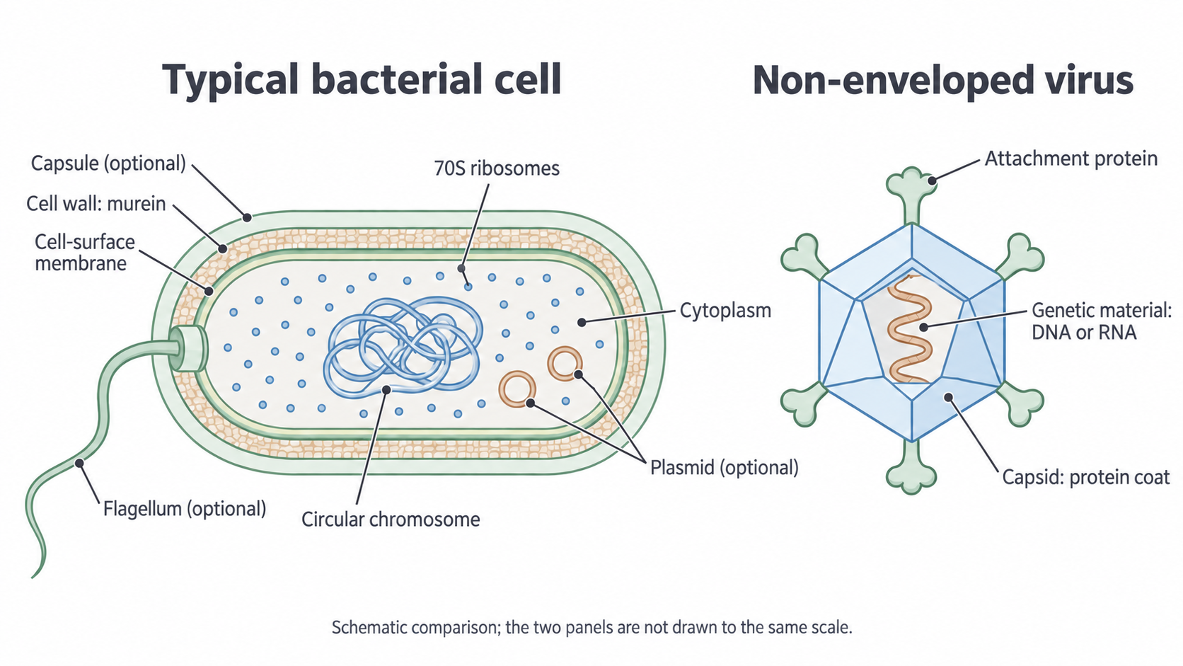
The large coiled DNA region is the chromosome, while the much smaller separate loops are plasmids. None of this DNA is enclosed by a nuclear membrane.
- Plasmids are smaller circular DNA molecules separate from the main chromosome, and a cell may have one or more. They can replicate independently and carry genes, sometimes including antibiotic-resistance genes.
- A capsule is a slimy outer layer surrounding the cell wall. It can protect the cell, reduce water loss or aid attachment to surfaces.
- One or more flagella may project from a bacterium. A flagellum is a long, thin structure whose rotation can propel the cell through liquid, so a bacterium without one cannot move in this way.
Virus structure and attachment
A virus has no cytoplasm and no ribosomes, so new virus particles can be made only inside a living host cell.
A virus particle is acellular: it is not a cell. The right-hand panel of the previous section’s diagram shows its three core parts:
- Genetic material, which is DNA or RNA
- A capsid, a coat made of protein that surrounds the genetic material
- Attachment proteins on the outside, which enable binding to a host cell
Some viruses also have a lipid envelope, but an envelope is not present in every virus and does not turn it into a cell.
Watch out
A capsid is not a capsule. A capsule surrounds a bacterial cell, whereas a capsid is the protein coat of a virus particle.
Why viruses are classed as non-living
Viruses are described as non-living because they lack cellular organisation and cannot carry out independent metabolism or protein synthesis. With no ribosomes, a virus depends on living host-cell machinery to make new virus components. So viruses do not grow and divide independently like bacterial cells.
Why a virus attaches to particular cells
Attachment depends on complementary shapes, as enzyme specificity does. An attachment protein binds to a receptor on the host-cell surface because its shape is complementary to that receptor. A cell without a suitable receptor cannot be entered by that route, which helps explain why a virus infects particular host species or cell types.
HIV, for example, has an attachment protein complementary to a receptor on helper T cells, a type of white blood cell, so these are among the cells it infects.
If a treatment changes the shape of an attachment protein, it may no longer bind its receptor. Fewer particles enter host cells, so fewer cells can produce new viruses.
Identifying cells and virus particles
One feature rarely settles an identification, so build your answer from several observations that agree.
A wall by itself does not establish that a cell is prokaryotic. Plants, algae and fungi also have walls, so its composition matters, and a wall alone does not show whether a nucleus is present. Circular DNA by itself is insufficient too, because mitochondria and chloroplasts contain circular DNA.
Combinations that identify a specimen
- Nucleic acid enclosed in a capsid, with no cytoplasm and no ribosomes, is consistent with a virus.
- Cytoplasm, ribosomes, a membrane and free circular DNA are consistent with a bacterium.
- A nucleus together with mitochondria supports eukaryotic identity, even if no chloroplasts are seen.
Absence requires cautious interpretation. “No nucleus visible in this image” can mean that the image or section missed it, rather than that none exists. Where no information is supplied about a feature, do not treat it as absent, and qualify your conclusion.
Three kinds of structure compared
| Observation | Typical bacterial cell | Typical eukaryotic cell | Virus particle |
|---|---|---|---|
| Genetic material | Circular chromosome free in cytoplasm | Protein-bound linear chromosomes in a nucleus | DNA or RNA enclosed in a capsid |
| Cytoplasm | Present | Present | Absent |
| Ribosomes | 70S | 80S in cytoplasm; 70S in mitochondria and chloroplasts | Absent |
| Membrane-bound organelles | Absent | Present | Absent |
| Surrounding structures | Cell-surface membrane; usually murein wall; sometimes capsule | Cell-surface membrane; wall in plants, algae and fungi | Protein capsid and attachment proteins; sometimes envelope |
In a comparison, state the same feature for both sides: prokaryotes have ribosomes, whereas viruses have none. One side alone can lose marks. Cover the table, then compare a bacterium with a virus from memory.