3.6.4.3 - Control of blood water potential
Blood water potential stays steady because the kidneys return most of the water they filter. Follow blood and filtrate through a nephron, then see how the medullary gradient and ADH set how much water stays in the body.
The kidney and the nephron
Your kidneys filter about 180 litres of fluid a day, yet you pass only 1.5 to 2 litres of urine.
About 99% of the filtered fluid returns to the blood, and changing how much water returns controls blood water potential.
Osmoregulation
Osmoregulation is the control of the water potential of the blood.
Losing water without an equivalent loss of solute lowers blood water potential (makes it more negative); gaining water raises it.
The path of filtrate
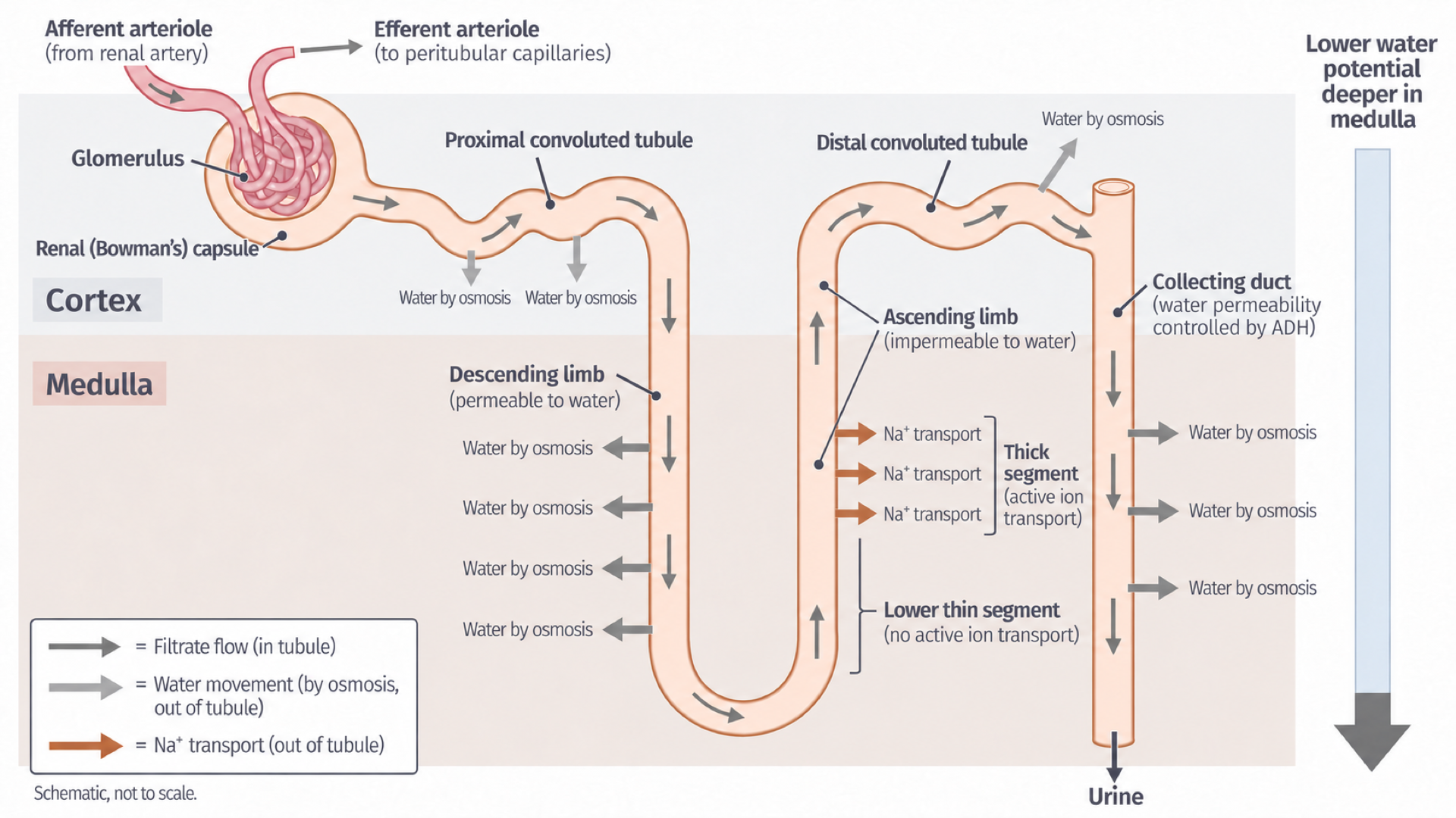
A nephron is a microscopic unit that filters blood and processes the filtrate. Filtrate passes through, in order:
- Renal capsule (Bowman's capsule), in the outer cortex, around a knot of capillaries called the glomerulus.
- Proximal convoluted tubule (PCT).
- Loop of Henle: the descending limb into the medulla, the bend, then the ascending limb.
- Distal convoluted tubule (DCT), back in the cortex.
- Collecting duct, down through the medulla towards the renal pelvis and ureter.
The path of blood
Blood follows a separate route: afferent arteriole → glomerular capillaries → efferent arteriole → capillaries around the tubules → renal vein. Reabsorbed substances pass from the tubule into tissue fluid and back into those capillaries. The glomerulus is not part of the tubule, and the efferent arteriole carries blood, not urine.
On the diagram, trace the dark filtrate arrows first. The paler arrows show water and sodium ions leaving across the tubule wall, not flowing along it.
[DIAGRAM: asset_name: Nephron and medullary gradient - rebuilt; asset_slug: Nephron and medullary gradient - rebuilt; recommended_method: retained_png; caption: Filtrate passes from renal capsule through PCT, loop of Henle and DCT into a collecting duct, water leaves water-permeable segments by osmosis.; description: Filtrate passes from renal capsule through PCT, loop of Henle and DCT into a collecting duct; water leaves water-permeable segments by osmosis.]
Formation of glomerular filtrate
Pressure forces fluid out of the glomerulus, and size, not usefulness, decides what gets through.
Ultrafiltration
Ultrafiltration is the pressure-driven filtration of water and small dissolved substances from glomerular blood into the renal capsule.
The efferent arteriole is narrower than the afferent arteriole, so it resists blood leaving the glomerulus. Hydrostatic pressure in the glomerular capillaries therefore stays high, forcing fluid across the filtration barrier.
Fluid crosses three layers, left to right on the diagram:
- Fenestrated endothelium: pores in the capillary wall, too small for blood cells.
- Basement membrane: a mesh too fine for most plasma proteins.
- Filtration slits: gaps between podocyte foot processes. Podocytes form the capsule's inner wall, and their foot processes wrap around the capillaries.
[DIAGRAM: asset_name: Glomerular filtration barrier - rebuilt; asset_slug: Glomerular filtration barrier - rebuilt; recommended_method: retained_png; caption: Pressure drives small solutes and water through endothelial pores, basement membrane and slits between podocyte foot processes into the renal capsule.; description: Pressure drives small solutes and water through endothelial pores, basement membrane and slits between podocyte foot processes into the renal capsule.]
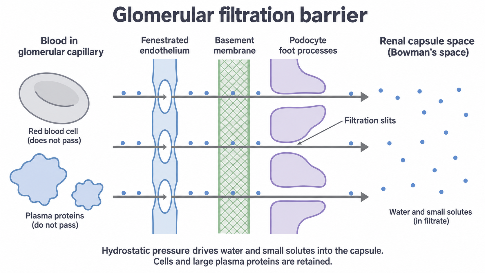
Filtrate normally contains water, glucose, amino acids, ions and urea. It is not urine yet: useful substances are reabsorbed later.
Exam tip
Asked why urine normally lacks protein, say plasma proteins are too large to pass through the basement membrane. Naming only "membrane", capillary, endothelium or glomerulus can lose the mark.
Filtration does not select by usefulness: urea and glucose both enter because they are small enough. Large proteins are held back by the barrier, not filtered and then removed. Pressure drives this movement, so it is not active transport by the capsule.
Reabsorbing glucose and water in the PCT
A sodium gradient, built by a pump on the blood side of the cell, draws glucose back out of the filtrate.
Selective reabsorption returns useful substances from filtrate to blood. Normally the PCT recovers essentially all filtered glucose, with much of the filtered water and ions. If filtered glucose exceeds transporter capacity, some stays in urine: the glycosuria of RP11.
How glucose returns to the blood
This is the ileum mechanism from 3.2.3, here in PCT epithelial cells:
- Sodium–potassium pumps in the blood-facing membrane use ATP to move sodium ions out into tissue fluid, from which they enter capillaries.
- This keeps intracellular sodium low, giving a gradient from filtrate to cell.
- At the lumen-facing membrane, sodium moves down that gradient through a sodium–glucose co-transporter, carrying glucose in, even against the glucose gradient.
- Glucose leaves through transport proteins in the blood-facing membrane by facilitated diffusion, into tissue fluid and blood.
ATP is used at the pump, not by the co-transporter.
How water follows
Moving solutes out of the filtrate lowers the water potential of the epithelial cells and tissue fluid relative to the filtrate. So water follows into tissue fluid and blood by osmosis; it is not pumped.
Exam tip
Water is reabsorbed in the PCT too. On a 2025 AQA nephron table, this was the tick most often missed.
Microvilli increase the lumen-facing surface for transport proteins, folds add area at the blood-facing membrane, and numerous mitochondria supply ATP for the pumps.
Continue this lesson.
You’re previewing NovaLearn without an account. Create a free account to read the rest.
Create free account