3.3.3 - Digestion and absorption
Starch, proteins and triglycerides are too large to cross cell-surface membranes. This lesson follows each through enzyme hydrolysis and absorption in the ileum of mammals, then shows how to investigate both.
Digestion as hydrolysis
A starch molecule is far too large to enter a cell, but the glucose units it is built from can be absorbed.
Digestion
Digestion is the hydrolysis of large biological molecules into smaller molecules that can be absorbed across cell membranes.
Proteins and triglycerides are also too large to be absorbed intact, so enzymes break them down step by step. Absorption follows: the products cross the epithelial cells lining the gut, then enter the blood or lymph.
Keep two ideas separate:
- Physical breakdown, such as chewing, breaks food into smaller pieces, which increases surface area.
- Chemical digestion uses enzymes to hydrolyse chemical bonds by adding water.
Digestive enzymes have active sites specific to particular substrates or bonds, so digestion often requires several enzymes:
| Food molecule | Bond hydrolysed | Main products absorbed |
|---|---|---|
| Starch and disaccharides | Glycosidic | Monosaccharides |
| Proteins | Peptide | Amino acids |
| Triglycerides | Ester | Fatty acids and monoglycerides |
Carbohydrate digestion
Starch digestion starts in your mouth, stalls in your stomach and finishes on the lining of the small intestine.
- In the mouth, salivary amylase begins the hydrolysis of starch.
- In the stomach, acid denatures this amylase, so starch digestion pauses.
- In the small intestine, alkaline secretions neutralise the stomach acid, so pancreatic amylase can continue, hydrolysing starch to maltose.
- Maltose is not absorbed intact through the monosaccharide transport pathways, so digestion continues at the epithelial surface of the small intestine, including the ileum.
Enzymes fixed to the cell surface
The enzymes there are membrane-bound disaccharidases. Membrane-bound means attached to the epithelial cell-surface membrane rather than released freely into the gut lumen.
| Enzyme | Substrate | Products |
|---|---|---|
| Maltase | Maltose | alpha-glucose + alpha-glucose |
| Sucrase | Sucrose | Glucose + fructose |
| Lactase | Lactose | Glucose + galactose |
In panel 1 of the diagram, amylase works in the gut lumen. In panel 2, maltase is fixed to the epithelial membrane, so glucose is produced exactly where absorption begins.
[DIAGRAM: asset_name: carbohydrate-digestion-v2; asset_slug: carbohydrate-digestion-v2; recommended_method: retained_png; caption: Amylase produces maltose, membrane-bound maltase hydrolyses each maltose to two glucose molecules outside the cell.; description: Amylase produces maltose; membrane-bound maltase hydrolyses each maltose to two glucose molecules outside the cell.]
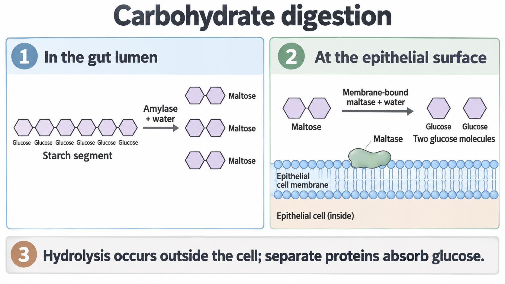
The active sites face the lumen, so hydrolysis happens outside the cell. Separate transport proteins then absorb the products: maltase does not carry maltose through the membrane.
Why too little lactase causes diarrhoea
Lactose intolerance shows what happens when too little lactase is produced. Lactose is not fully hydrolysed in the ileum and reaches the large intestine, where microorganisms act on it. The soluble products lower the water potential of the colon contents, so water enters by osmosis and diarrhoea can result.
Lipid digestion
Lipase dissolves in water but fat does not, so the enzyme can only reach the surface of a fat droplet.
Lipids are not soluble in the largely aqueous gut contents, so they form large droplets, with most of their triglyceride out of reach inside.
How bile salts speed up lipase
Bile salts, produced by the liver and released into the small intestine, carry out emulsification:
- Gut mixing breaks large droplets into smaller ones.
- Each bile salt has a lipid-soluble side and a water-soluble side, so bile salts coat a droplet with their water-soluble sides facing outwards.
- Coated droplets cannot easily rejoin, so they stay small.
- Many small droplets have a much larger total surface area, so more triglyceride is within reach of lipase and hydrolysis is faster.
Emulsification is a physical change: it does not hydrolyse ester bonds or create fatty acids, and bile salts are not enzymes.
From lipase to micelles
Lipase hydrolyses triglycerides to fatty acids and monoglycerides.
Watch out
Students often write “glycerol and fatty acids” here, because hydrolysing a triglyceride in 3.1.3 released glycerol. In the gut, lipase usually breaks only two of the three ester bonds, giving two fatty acids and one monoglyceride: glycerol with one fatty acid still attached.
These products stay associated with bile salts in micelles, which carry them to the epithelial surface. The diagram shows the order: emulsification (1), lipase action (2), then a micelle heading for the ileum surface (3).
[DIAGRAM: asset_name: 3.3 - Digestion and absorption - Diagram 2; asset_slug: 3.3 - Digestion and absorption - Diagram 2; recommended_method: retained_png; caption: Bile salts assist emulsification, lipase produces fatty acids and monoglycerides, which associate with bile salts in micelles.; description: Bile salts assist emulsification; lipase produces fatty acids and monoglycerides, which associate with bile salts in micelles.]
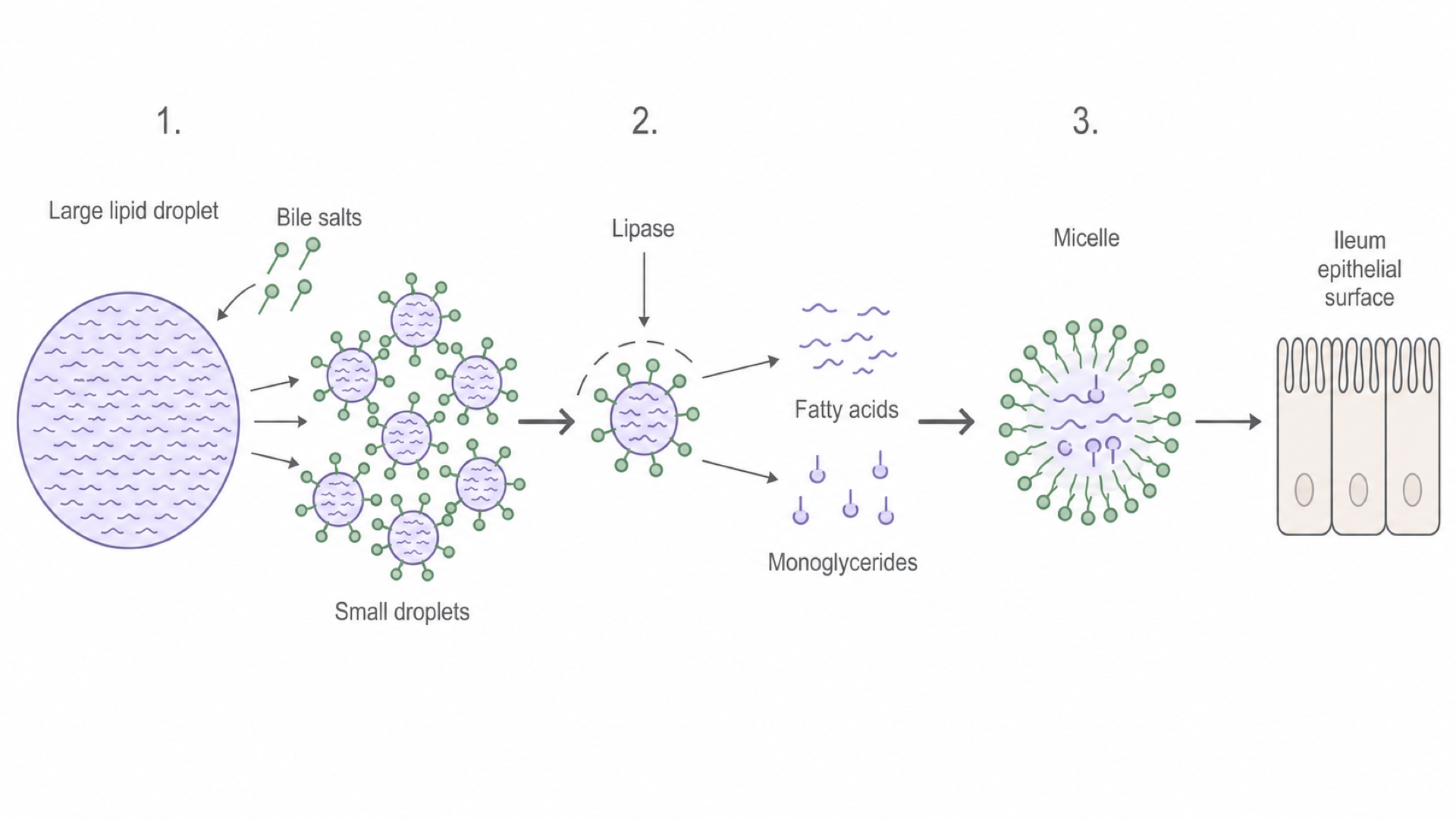
Emulsification does not form micelles: they form afterwards, from the products of lipase.
Exam tip
Give emulsification and micelles a point each. For the rate, write “faster hydrolysis by lipase”: AQA’s June 2025 mark scheme ignored “digestion” and “breakdown” there.
| Emulsified droplet | Micelle | |
|---|---|---|
| Stage | Before lipase acts | After lipase acts |
| Contains | Triglycerides | Fatty acids and monoglycerides |
| Effect | More surface area, so faster hydrolysis | Carries products to the epithelial surface |
Cover the table, then state from memory what each one does.
Protein digestion
Three kinds of enzyme dismantle a protein in turn, and the first speeds up the second.
Protein digestion uses enzymes called peptidases or proteases, which hydrolyse peptide bonds:
- Endopeptidases hydrolyse peptide bonds within the middle of a polypeptide chain, producing many shorter fragments.
- Exopeptidases hydrolyse peptide bonds at the ends of those fragments, releasing amino acids and dipeptides.
- Membrane-bound dipeptidases at the epithelial surface of the ileum hydrolyse dipeptides into amino acids, ready for absorption.
Exopeptidases act at chain ends, so one long protein offers few sites. Each endopeptidase cut creates two new ends, so the fragments give exopeptidases many more sites and hydrolysis is much faster.
The diagram shows each enzyme as a separate example, so the chains do not carry over between panels. In panel 1, one chain with two ends becomes two chains with four ends.
[DIAGRAM: asset_name: protein-digestion-v2; asset_slug: protein-digestion-v2; recommended_method: retained_png; caption: Separate examples compare internal peptide-bond hydrolysis, terminal hydrolysis and the splitting of a dipeptide into two amino acids.; description: Separate examples compare internal peptide-bond hydrolysis, terminal hydrolysis and the splitting of a dipeptide into two amino acids.]
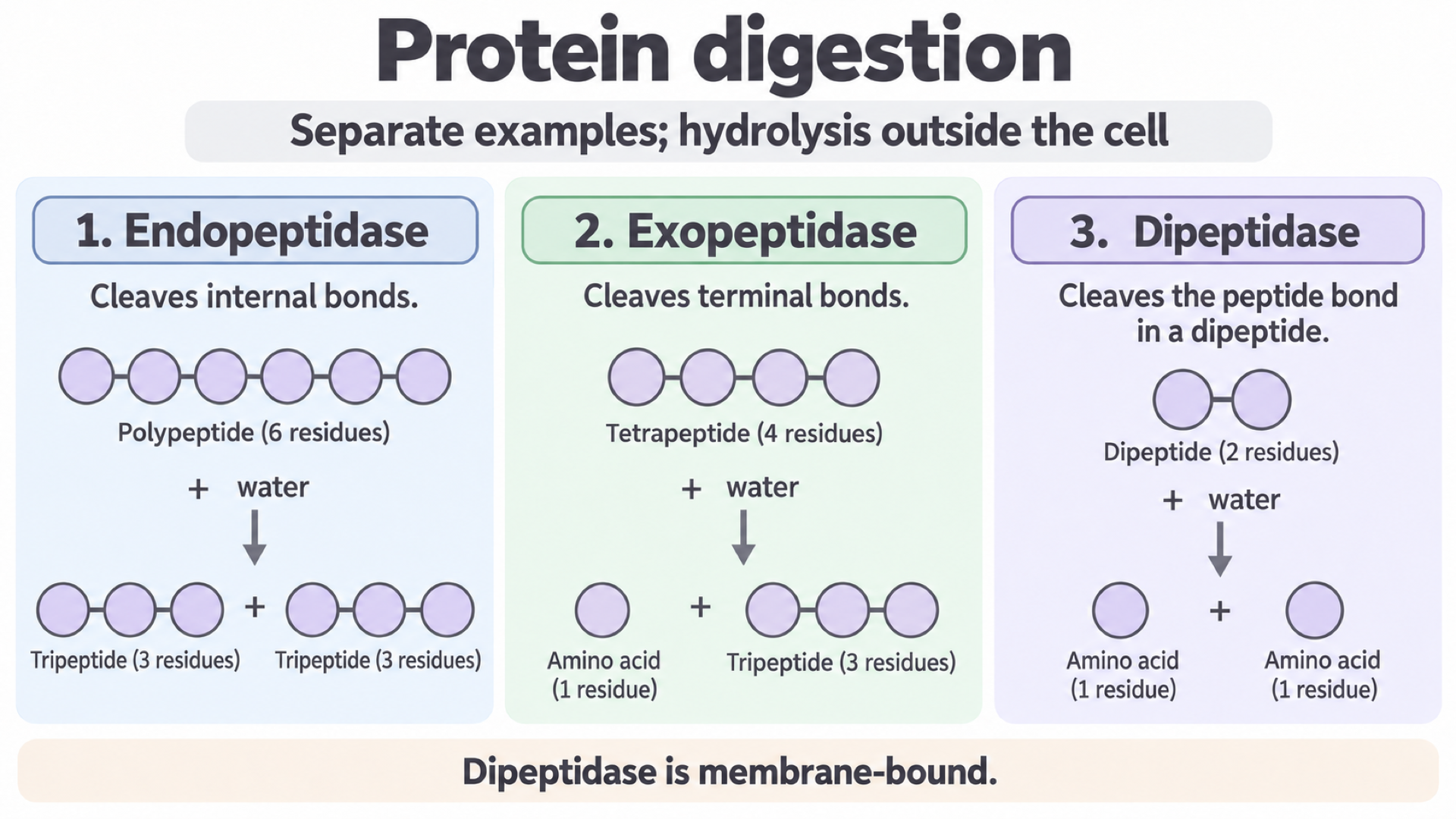
A dipeptidase, like maltase, is attached to the membrane but is not a transport protein: separate proteins absorb the amino acids.
The ileum and co-transport
A molecule in the gut is still outside your body until it crosses the epithelial cells lining the ileum.
How the ileum speeds up absorption
- The ileum has many villi, multicellular projections of its wall, which increase surface area.
- Each epithelial cell has microvilli, folds of its cell-surface membrane, which increase surface area further.
- The epithelium is thin, so the diffusion pathway is short.
- A rich capillary network carries absorbed molecules away, which helps maintain concentration gradients.
Exam tip
Asked what microvilli are, write “folds of the cell-surface membrane”. AQA’s June 2024 mark scheme rejected “hairs”.
The apical membrane of an epithelial cell faces the lumen and carries the microvilli. The basal membrane (also called the basolateral membrane) faces the tissue fluid and blood capillaries.
How sodium ions drive co-transport
Glucose and galactose are absorbed by co-transport with sodium ions, which you met in 3.1.8 and 3.2.3, and many amino acids use sodium-dependent co-transporters too. Not every monosaccharide does: fructose uses facilitated diffusion at the apical membrane.
The diagram follows glucose. Start at the pump, cross to the co-transporter, then finish at the carrier at bottom right.
[DIAGRAM: asset_name: 3.3 - Digestion and absorption - Diagram 4; asset_slug: 3.3 - Digestion and absorption - Diagram 4; recommended_method: retained_png; caption: A basolateral sodium-potassium pump maintains the sodium gradient that drives apical glucose co-transport, glucose exits by facilitated diffusion.; description: A basolateral sodium-potassium pump maintains the sodium gradient that drives apical glucose co-transport; glucose exits by facilitated diffusion.]
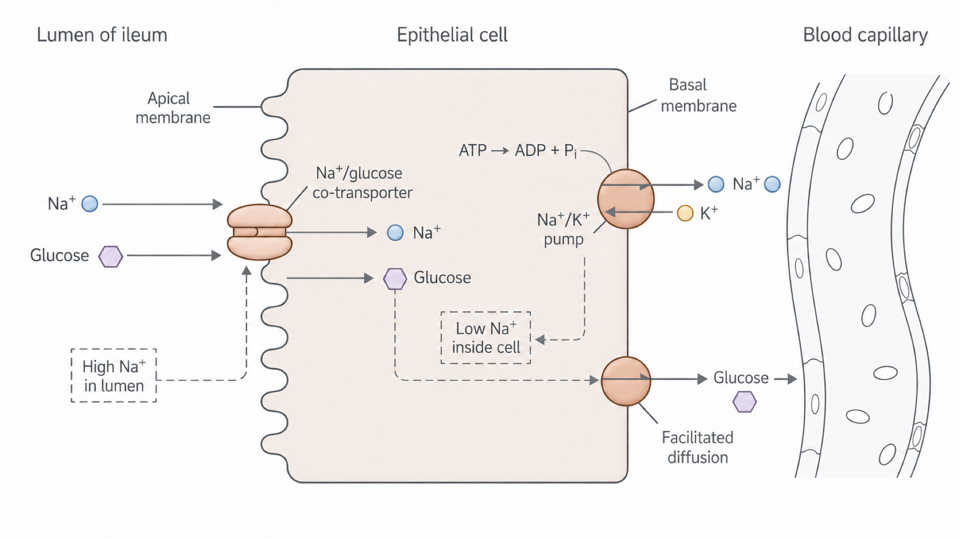
- On the basal membrane, the sodium-potassium pump uses ATP to move sodium ions out into the tissue fluid and potassium ions into the cell.
- This keeps the sodium ion concentration lower inside the cell than in the lumen.
- Sodium ions therefore move back in down their concentration gradient, through a co-transporter in the apical membrane.
- Glucose or an amino acid is carried in with the sodium ion, even if it is moving against its own concentration gradient.
- The nutrient is now more concentrated in the cell than in the blood, so it leaves by facilitated diffusion through carrier proteins in the basal membrane. Blood flow carries it away, which helps maintain this gradient.
Continue this lesson.
You’re previewing NovaLearn without an account. Create a free account to read the rest.
Create free account