3.6.3 - Skeletal muscle
Skeletal muscles are the effectors that turn nerve impulses into movement. Follow one movement down to myosin heads pulling on actin, then see why some fibres tire sooner and how to study muscle.
Antagonistic pairs and the skeleton
A muscle can pull but not push, so straightening a bent arm takes a different muscle from the one that bent it.
A skeletal muscle generates tension, and tendons transmit that pull to bone. The skeleton is relatively incompressible, so bones act as rigid levers and the tension moves them around a joint. Ligaments hold bones together at joints.
Antagonistic muscles
Muscles that produce opposing movements at a joint: as one contracts, the other relaxes.
In the diagram, green marks the contracting muscle. In flexion, the biceps contracts while the triceps relaxes and is stretched. In extension the roles swap. Relaxing the biceps only removes its pull: the triceps pulls the forearm back.
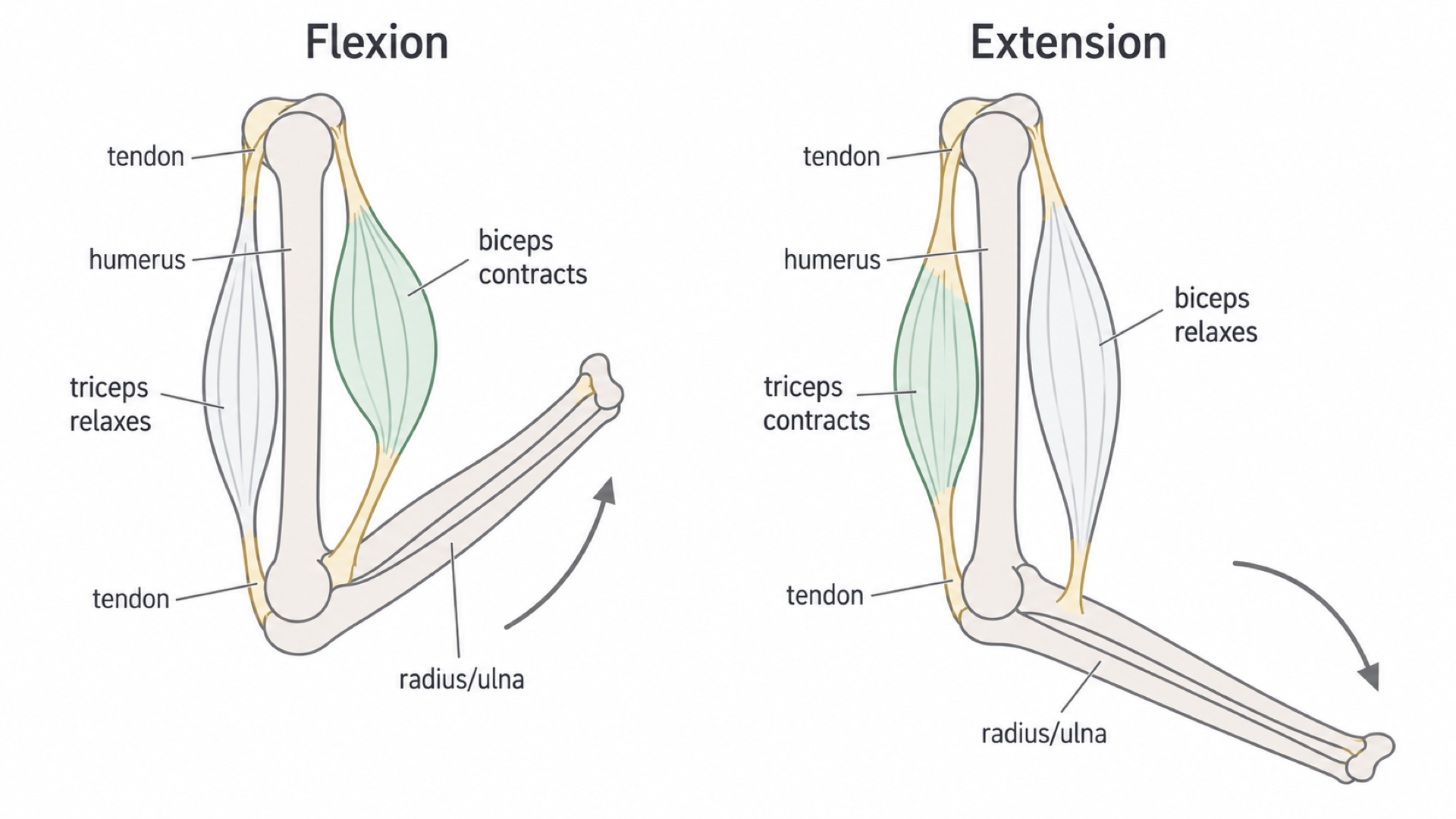
Contraction means generating tension, not necessarily shortening: a muscle holding a load contracts with almost no change in length.
Muscle structure and the sarcomere
Zoom in from a whole muscle to its protein filaments, and the stripes of muscle explain how it shortens.
Skeletal muscle is built in levels:
- A whole muscle contains bundles called fascicles.
- Each fascicle contains many muscle fibres, long multinucleate cells enclosed by a sarcolemma.
- A fibre's cytoplasm, the sarcoplasm, contains mitochondria and many parallel myofibrils. The sarcoplasmic reticulum surrounds them and stores calcium ions.
- A myofibril is a chain of repeating contractile units, sarcomeres, containing thin actin filaments and thick myosin filaments.
The bands of a sarcomere
One sarcomere extends from one Z line to the next. Actin is anchored to the Z lines and extends towards the centre. Myosin lies centrally, with heads projecting towards the actin. Tropomyosin lies along actin and covers the myosin-binding sites at rest.
| Region | What it contains | Change during shortening |
|---|---|---|
| A band | The full length of the thick myosin filaments, including overlap | Width unchanged |
| I band | Actin without myosin; a full I band spans a Z line between neighbouring sarcomeres | Narrows |
| H zone | Myosin without actin in the centre of the A band | Narrows and can disappear |
| Sarcomere | Z line to Z line | Shortens |
Dark A bands contain thick myosin, and light I bands contain only thin actin. Neighbouring myofibrils line up, so these stripes run across the whole fibre, giving skeletal muscle its striated look.
In a transverse section of a myofibril, small dots alone mean actin (an I band), large dots alone mean myosin (the H zone), and both together mean the overlap region of the A band.
What changes when a sarcomere shortens
During contraction, actin slides farther between the myosin filaments, bringing the Z lines closer. Neither filament shortens. More overlap reduces the actin-only and myosin-only regions. Because the filaments keep their lengths, the H zone and the full I band each narrow by exactly as much as the sarcomere shortens.
The diagram shows one sarcomere relaxed and contracted, at the same scale: the myosin ends stay aligned while the Z lines move in.
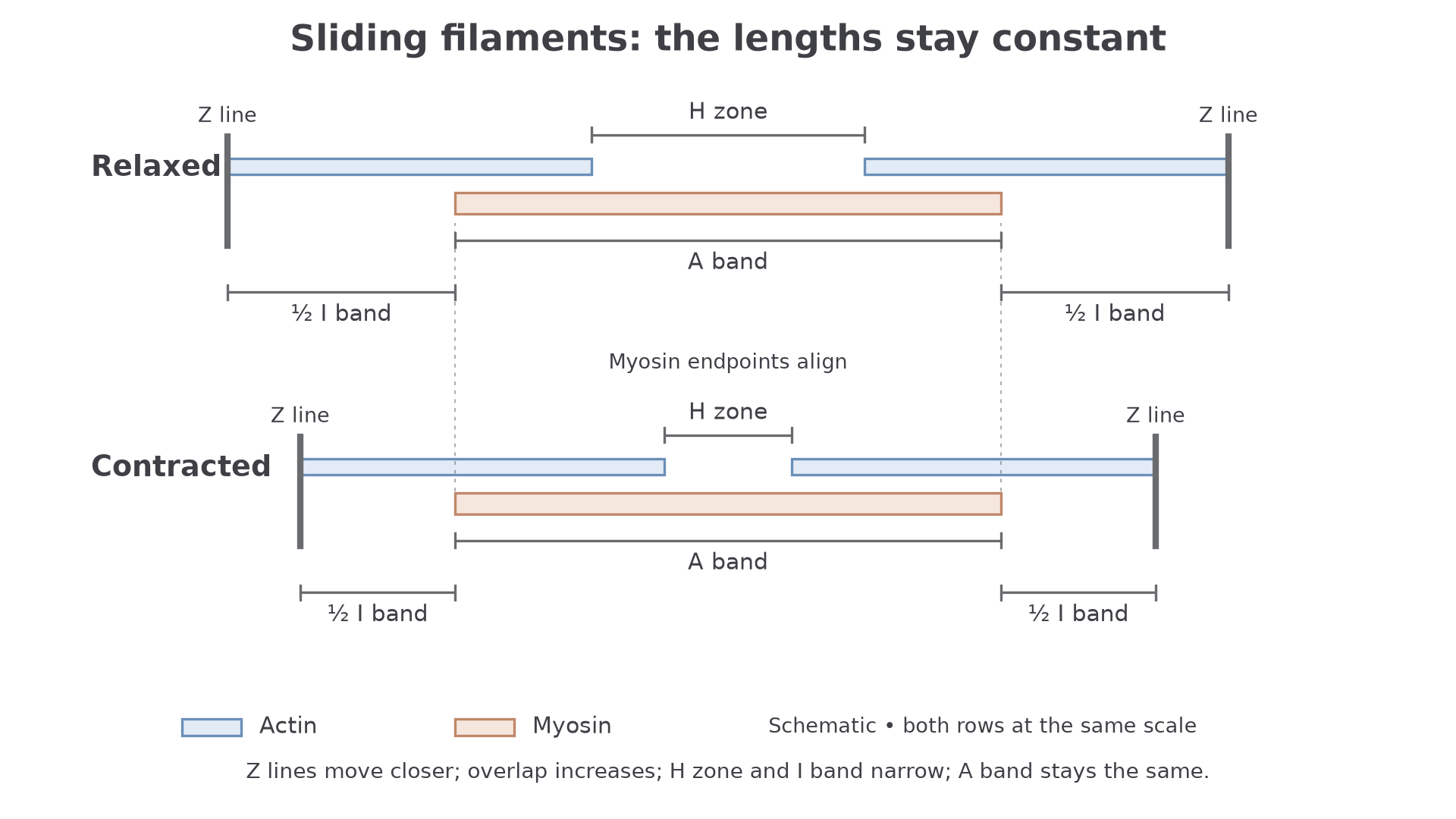
The actin-only region at each end is half of an I band, so do not label a whole thin filament as an I band.
Worked example: myofibril length
A myofibril has 20 sarcomeres, each 2.5 µm long. Each shortens by 20%. Give the new length in millimetres, in standard form.
Plan: find the total, keep 80% of it, then convert units.
- µm
- A 20% decrease leaves 80%: µm
- mm
Answer: .
Check: 10 µm is the decrease, not the new length, a slip AQA reported in 2024. The power of ten was essential for full marks.
So a band set by a filament's length stays constant, and a band set by the gap between filament ends narrows.
From motor neurone to calcium release
One impulse at the surface of a fibre ends with calcium ions flooding the myofibrils deep inside it.
You met the neuromuscular junction in 3.6.2.2, where a motor-neurone terminal meets the sarcolemma. Each fibre is activated through its own junction. A motor neurone and the fibres it supplies form a motor unit, and recruiting more motor units increases a muscle's force.
The sequence is:
- An action potential opens voltage-gated calcium channels in the motor-neurone terminal, so calcium ions enter.
- This triggers acetylcholine release by exocytosis.
- Acetylcholine diffuses across the cleft and binds to receptors on the sarcolemma.
- Channels open, so sodium ions enter, depolarising the sarcolemma and normally triggering a muscle action potential.
- The action potential spreads along the sarcolemma and down T-tubules, inward extensions of that membrane.
- Calcium-release channels open in the sarcoplasmic reticulum, so calcium ions move from this store into the sarcoplasm around the myofibrils.
Exam tip
Name the membrane and the ion. In 2024, many students wrote “muscle” instead of sarcolemma, and “sodium ion channels opening” alone was not enough: say sodium ions enter.
T-tubules carry the signal inward rapidly, so deep myofibrils get calcium ions almost as soon as those near the surface, and they contract together.
Keep the two calcium movements apart. Calcium ions from outside the cell enter the motor-neurone terminal and trigger transmitter release. Calcium ions stored in the muscle's sarcoplasmic reticulum enter the sarcoplasm and allow contraction.
The sliding filament mechanism
Myosin heads pull actin in short strokes, and calcium ions and ATP control when each stroke can happen.
Contraction depends on repeated actinomyosin cross-bridges, temporary links between myosin heads and binding sites on actin.
At rest, tropomyosin blocks these sites. When the calcium ion concentration in the sarcoplasm rises, regulatory proteins change shape and tropomyosin moves away, exposing the sites. Calcium ions are not an energy source and do not pull actin.
The cross-bridge cycle
While the sites stay exposed, each myosin head repeats the four steps in the diagram.
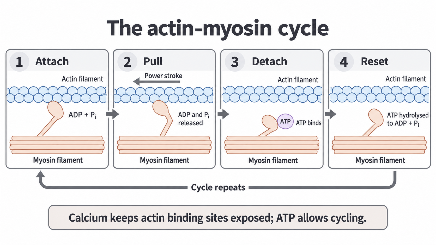
- Attach. An energised myosin head carrying ADP and inorganic phosphate binds to an exposed site, forming a cross-bridge.
- Pull. The head pivots in a power stroke, using energy stored at step 4 to pull actin towards the sarcomere centre. ADP and phosphate are released.
- Detach. A new ATP molecule binds to the head, causing it to detach from actin.
- Reset. Myosin's ATPase hydrolyses ATP to ADP and inorganic phosphate. The released energy resets the head, so it can bind farther along the actin.
Heads at opposite ends of the thick filament pull the two sets of actin towards the centre, so overlap increases. Many heads work asynchronously, so tension continues as individual heads detach.
Watch out
ATP binding causes detachment; ATP hydrolysis resets the head. AQA listed “binding to actin via condensation and detaching via hydrolysis” as a common significant error in 2025.
Relaxation also needs ATP
When stimulation ends, calcium ions are actively pumped back into the sarcoplasmic reticulum using ATP. Tropomyosin covers the sites again, so no new cross-bridges form. The antagonist's pull can then lengthen the relaxed muscle.
Without ATP, attached heads cannot detach normally, which explains the stiffness of rigor mortis.
Continue this lesson.
You’re previewing NovaLearn without an account. Create a free account to read the rest.
Create free account