3.8.4.2 - DNA probes and genetic screening
One changed base in an allele can cause a heritable condition or make a standard drug dose harmful. This lesson shows how labelled DNA probes find such differences between people, and how the results guide genetic counselling and personalised medicine.
DNA probes
A probe turns one allele's base sequence into a signal you can detect.
Its base sequence finds the target, and its label shows where it has bound. The label is also called a marker.
DNA Probe
A DNA probe is a short, single-stranded fragment of DNA that carries a complementary base sequence to a specific target sequence and is attached to a detectable label, such as a radioactive isotope or fluorescent dye.
How a probe tells alleles apart
A probe is made exactly complementary to the allele being sought, because adenine pairs with thymine and cytosine pairs with guanine.
In the diagram, follow the blue probe from left to right: each base sits opposite its partner in the target strand, and the dotted lines are the hydrogen bonds between them. The box at the end is the label.
[DIAGRAM: asset_name: 8.4.2 - Differences in DNA between individuals of the same species can be exploited for identification and diagnosis of heritable conditions - Diagram 1; asset_slug: 8.4.2 - Differences in DNA between individuals of the same species can be exploited for identification and diagnosis of heritable conditions - Diagram 1; recommended_method: retained_png; caption: A labelled single-stranded DNA probe hybridises to its complementary target sequence.; description: A labelled single-stranded DNA probe hybridises to its complementary target sequence.]
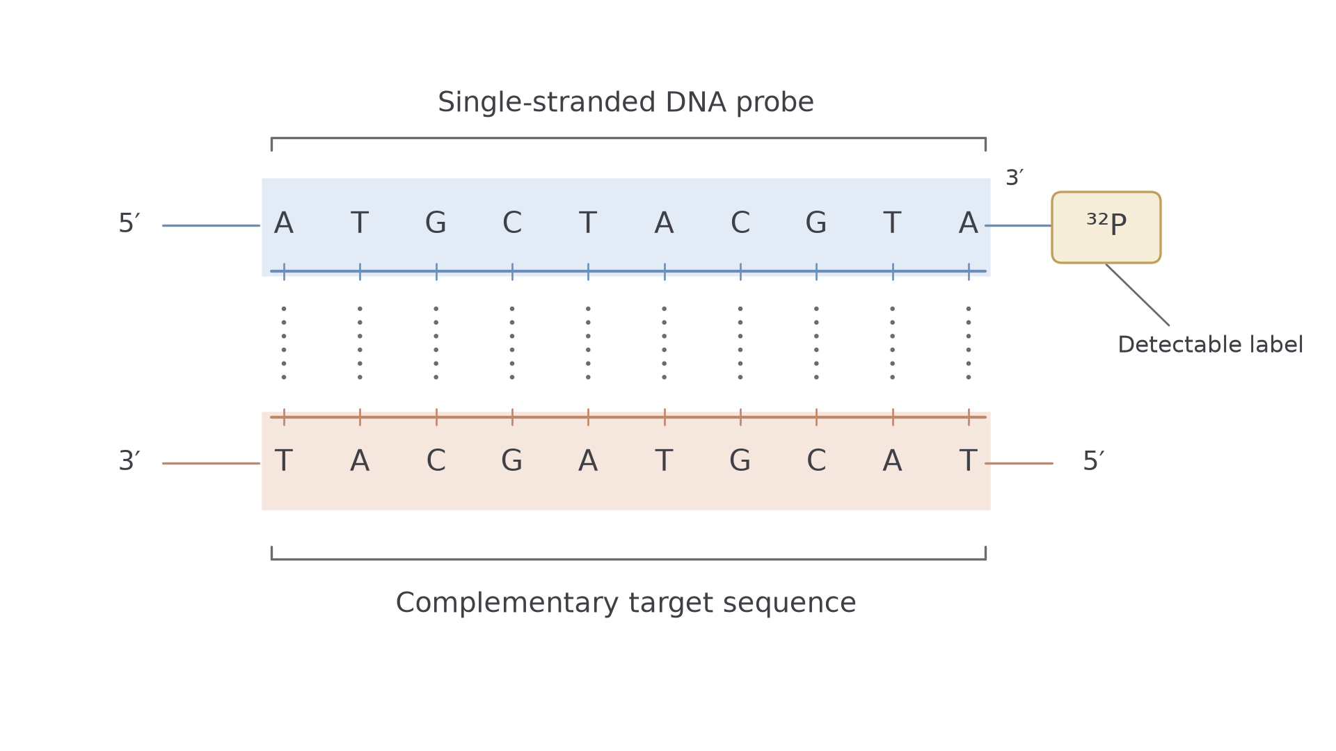
A mismatch means fewer hydrogen bonds hold the probe to the DNA, so it is removed more easily during washing. With suitable temperature and washing conditions, an exact match stays bound while a mismatched probe comes off. This is how a probe can tell apart two alleles that differ by a single base.
Exam tip
"What is a DNA probe?" was a 2-mark question in 2025. The marks went to a single strand of DNA and to bases complementary to the allele. Writing only "a short section of DNA", or only about labels and uses, can lose both.
Making and labelling a probe
Before a probe can be made, the base sequence of the target allele must be known. It comes from DNA sequencing or, for many common disorders, from genetic libraries that store allele sequences.
A short probe can then be synthesised chemically, as in a gene machine (3.8.4.1), with the label at one end or incorporated within it. If a sample holds too little target DNA, PCR can amplify it first.
Two main types of label make probes detectable:
- Radioactively labelled probes contain nucleotides with a radioactive isotope, typically phosphorus-32 (). X-ray film is placed over the sample, and the radiation exposes the film where the probe has bound. This is called autoradiography.
- Fluorescently labelled probes carry a fluorescent dye. Light of a specific wavelength, often ultraviolet, makes the bound dye fluoresce, and a specialised microscope shows where.
Locating an allele by hybridisation
Hybridisation lets a probe find its allele, and washing means any signal left comes from bound probe.
DNA Hybridisation
DNA hybridisation is the process by which a single-stranded DNA probe binds to a complementary single-stranded DNA sequence through complementary base pairing, forming a double-stranded hybrid molecule.
In double-stranded DNA the bases are already paired, so a probe has nothing to pair with. Heating denatures the DNA: the hydrogen bonds between complementary bases break and the two strands separate, exposing the bases. When the mixture cools with the probe present, the probe anneals (binds) to any complementary sequence, forming a hybrid molecule.
Steps to locate a specific allele
These steps locate a mutant allele that causes a genetic disorder:
- Identify the target sequence. DNA sequencing or a genetic library gives the base sequence of the mutant allele.
- Construct the probe. A short DNA fragment complementary to that allele is synthesised and labelled with a radioactive isotope or fluorescent dye.
- Obtain enough target DNA. DNA is extracted from the patient. If there is too little, the relevant region is amplified using suitable primers.
- Prepare the patient's DNA. Heating separates it into single strands, so its bases are free to pair with the probe.
- Add the probe. The single-stranded DNA is cooled in a mixture containing the labelled probes.
- Hybridisation. If the mutant allele is present, the probe binds to the complementary sequence.
- Wash away unbound probes. The patient's DNA is held on a solid surface, such as a membrane, so it stays behind. Washing matters because leftover probe would give a signal whether or not the allele is present.
- Detection. For a fluorescent probe, light of a specific wavelength makes bound probes fluoresce. For a radioactive probe, X-ray film is exposed. Either way, the signal shows where the probe has bound.
What a signal does and doesn't show
A signal means the target sequence is in the sample, as long as the controls behave as expected. It doesn't show whether the person has one copy or two.
Watch out
No signal doesn't prove the allele is absent. Failed extraction, too little DNA or unsuitable conditions also give no signal, so the safe conclusion is "target not detected".
Controls show which conclusion you can trust:
- A processing control is a second probe for a DNA sequence present in every valid human sample. If it gives no signal either, the test failed for that sample and must be repeated.
- A known target-positive sample checks that detection works.
- A known non-target sample checks specificity.
- A reagent blank (all the reagents but no DNA) checks contamination.
Genetic screening
A screening result can give a genotype, a probability or a raised risk, and each means something different.
Genetic Screening
Genetic screening is the process of testing individuals for the presence of specific alleles associated with genetic disorders, disease susceptibility, or altered drug responses, typically using DNA probes and hybridisation.
Screening is particularly important for people with a family history of a genetic disorder. It shows whether they carry the allele and allows calculation of the probability that their children could be affected.
Testing for many alleles at once
Hundreds of different DNA probes can be fixed in an array on a single glass slide, known as a DNA microarray. Each probe has a known position, so one sample can be tested for many genetic conditions at once.
In the array design shown, the probes are unlabelled. In panel 1, patient DNA is labelled, made single-stranded and added to the slide. The label is on the patient DNA here, not the probe, but it does the same job: it shows where hybridisation has happened.
[DIAGRAM: asset_name: 8.4.2 - Differences in DNA between individuals of the same species can be exploited for identification and diagnosis of heritable conditions - Diagram 2; asset_slug: 8.4.2 - Differences in DNA between individuals of the same species can be exploited for identification and diagnosis of heritable conditions - Diagram 2; recommended_method: retained_png; caption: Labelled patient DNA binds to complementary fixed probes, signal positions identify detected targets.; description: Labelled patient DNA binds to complementary fixed probes; signal positions identify detected targets.]
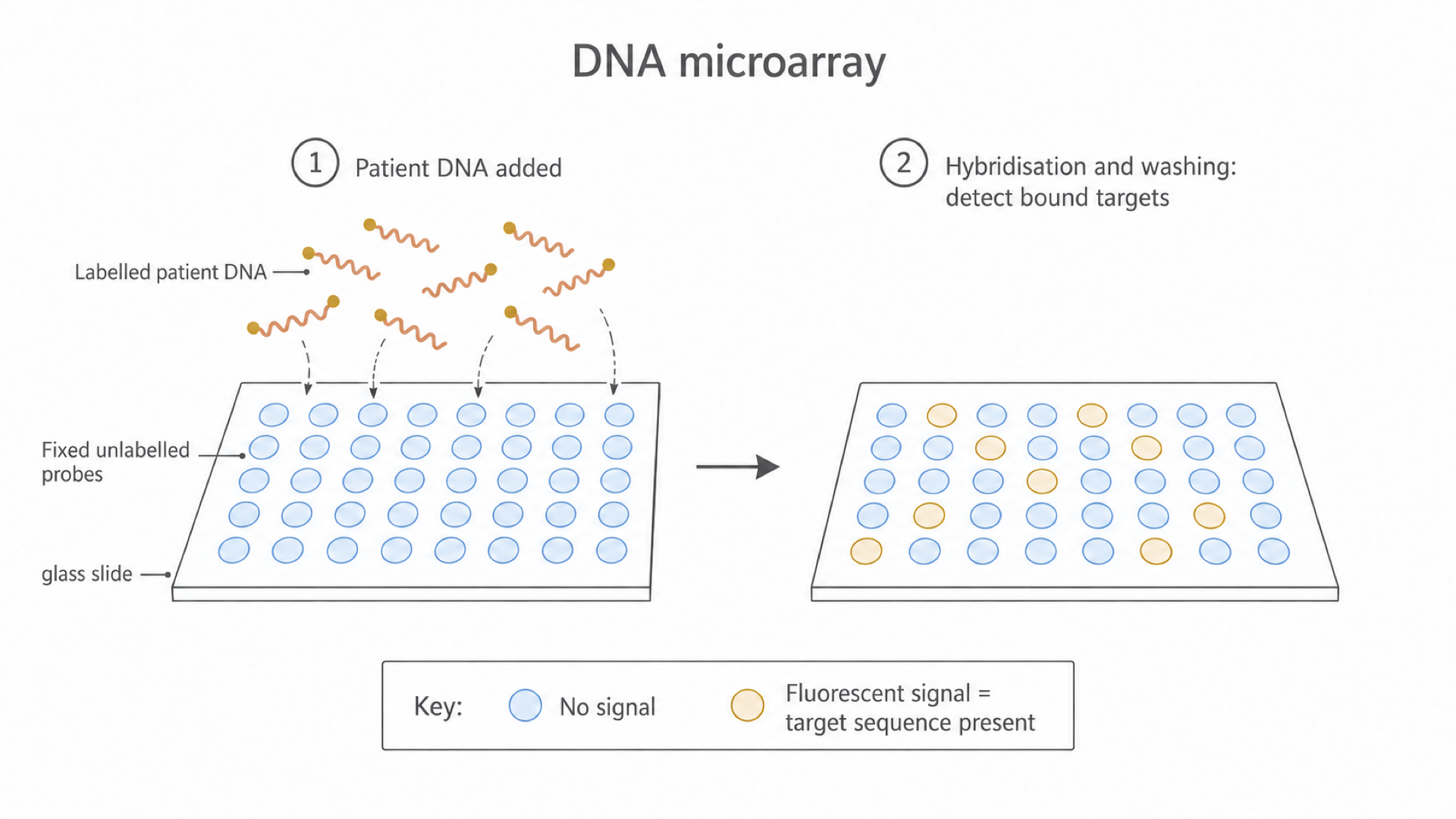
In panel 2, complementary sequences have hybridised and unbound material has been washed away. Orange spots fluoresce, and each position tells you which target sequence is present.
Blue spots show no detectable hybridisation, though controls are needed before inferring that a target is absent. The blue is only drawn in: fixed probes don't fluoresce before a sample is added.
Risk alleles are not diagnoses
Some alleles raise risk rather than cause a condition with certainty. Some inherited faulty alleles of the tumour suppressor genes BRCA1 and BRCA2 increase the risk of breast and ovarian cancers.
As you saw in 3.8.2.3, both copies of many tumour suppressor genes must be lost or inactivated in a cell, so someone inheriting one faulty copy needs fewer further changes. Such an allele has incomplete penetrance: not everyone who has it develops the condition, so a positive result shows raised risk, not a certain outcome.
Screening lets people make informed decisions about increased surveillance, avoiding known mutagens, lifestyle changes or preventative treatment, so that any cancer is detected early, when treatment is most effective.
Continue this lesson.
You’re previewing NovaLearn without an account. Create a free account to read the rest.
Create free account