3.4.3 - Genetic diversity from mutation and meiosis
Two human parents could produce about 70 trillion chromosome combinations in their children, before crossing over adds more. Mutation creates new alleles; meiosis and random fertilisation reshuffle existing ones.
Gene mutations and mutagens
One changed base can leave a protein untouched or stop it working, depending on how the code is read.
Gene mutation
A gene mutation is a change in the DNA base sequence of a gene.
Alleles differ in base sequence, so a mutation can create a new allele. Mutations can arise spontaneously when DNA is replicated. They are not directed toward what an organism needs, and their effect may be harmful, neutral or beneficial.
Mutagenic agents, such as ultraviolet radiation, ionising radiation and some chemicals, increase the rate of mutation.
Base substitution and the degenerate code
A base substitution replaces one base with another, normally changing one codon without shifting the reading frame. The new codon may specify:
- The same amino acid, because the code is degenerate. Primary structure is unchanged, so folding and function need not change. Check any code table: assuming a protein always changes can lose marks.
- A different amino acid. Its R group can form different bonds, which may change tertiary structure and function, though the protein need not become non-functional.
- A stop signal, which shortens the polypeptide.
Base deletion and the reading frame
A base deletion removes a nucleotide. Deleting one base within the translated sequence shifts the reading frame, regrouping downstream bases into different codons, so many later amino acids may change and a premature stop may occur. Deleting a complete triplet does not shift the frame.
| Coding DNA example (5′ to 3′) | Reading in the original frame |
|---|---|
| Original: ATG AAA GGC TTT | Four complete triplets |
| Substitute first A of AAA with G: ATG GAA GGC TTT | One triplet changed; later boundaries unchanged |
| Delete first A of AAA: ATG AAG GCT TT… | All later boundaries shift; the final two bases need the next base to complete a triplet |
A change outside the codons can still matter, for example at a splice site, where an intron is removed.
Meiosis and chromosome-content diagrams
One round of DNA replication and two divisions turn one diploid cell into four haploid cells.
A diploid cell has two sets of chromosomes; a haploid cell has one set.
Meiosis
Meiosis is a division sequence in which one diploid cell usually produces four haploid daughter cells.
What each division separates
- DNA replicates once before meiosis I. Each chromosome becomes two sister chromatids joined at the centromere, so DNA content doubles but chromosome number does not.
- Meiosis I separates homologous chromosomes. Homologues, which carry the same genes at corresponding loci, pair and move to opposite poles. Each nucleus receives one homologue from every pair, so it is haploid, though each chromosome still has two chromatids.
- Meiosis II separates sister chromatids, with no further DNA replication. Each separated chromatid now counts as a chromosome, so chromosome number does not halve again but DNA content per cell does.
In the diagram, colour shows parental origin and length shows chromosome type. Follow the long blue chromosome from left to right.
[DIAGRAM: asset_name: meiosis-lineage-v2; asset_slug: meiosis-lineage-v2; recommended_method: local_png; caption: A diploid cell with two homologous chromosome pairs produces two cells after meiosis I and four haploid products after meiosis II, crossing over is omitted.; description: A diploid cell with two homologous chromosome pairs produces two cells after meiosis I and four haploid products after meiosis II; crossing over is omitted.]
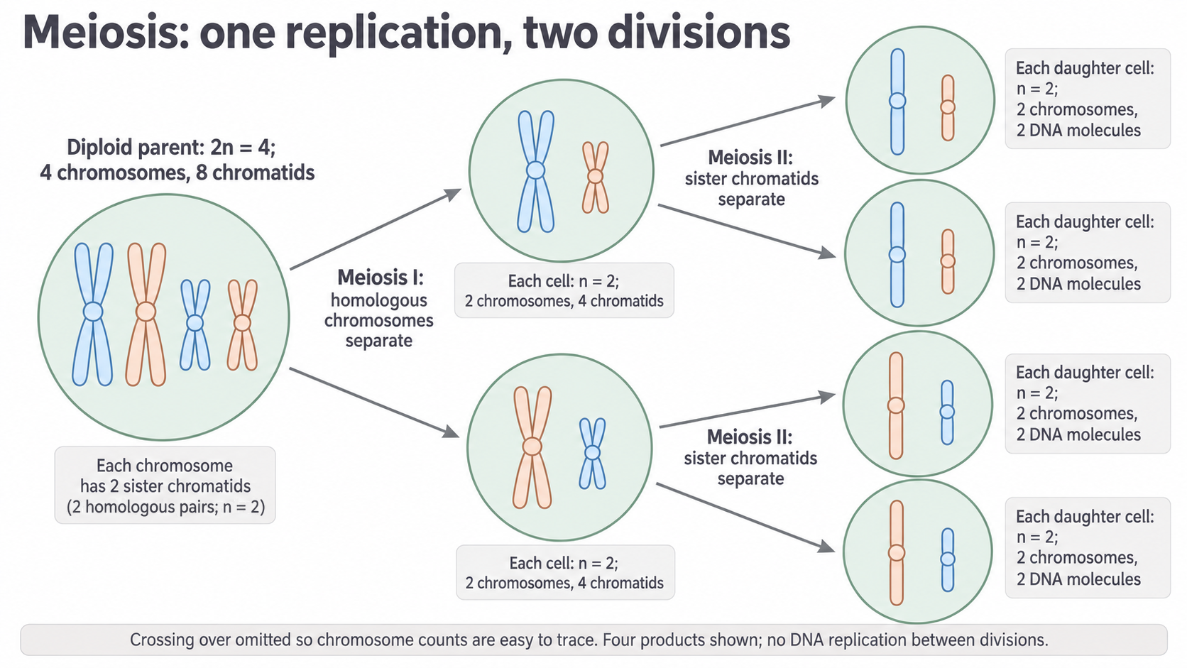
The middle cells hold two-chromatid X shapes; the final cells hold single rods, because sisters have separated. Each final cell needs one long and one short chromosome, not any two. The products of each meiosis-I cell match here only because crossing over is omitted; real meiosis can make them genetically different.
| Stage for a cell with | Cells | Chromosomes per cell | DNA molecules per cell |
|---|---|---|---|
| Before DNA replication | 1 | 4 | 4 |
| After replication, before meiosis I | 1 | 4 | 8 |
| End of meiosis I | 2 | 2 | 4 |
| End of meiosis II | 4 | 2 | 2 |
Completing a chromosome-content diagram
Count centromeres: joined sisters count as one chromosome until their centromeres separate.
- Identify homologous pairs from lengths and loci.
- Send one homologue from each pair to each meiosis-I cell, keeping sisters together.
- Draw one single chromatid from each chromosome in each meiosis-II product.
Exam tip
After meiosis I, draw one chromosome of each type, still with two chromatids. Keeping every chromosome or drawing single chromatids is a common error.
Observing prepared slides
Prepared anther tissue, from the pollen-producing part of a flower, or suitable animal reproductive tissue can contain cells in meiosis.
- Start the microscope at low power, which shows a wider area, to locate intact dividing cells. Centre them, then increase magnification.
- Use fine focus and adjust illumination for contrast, so chromosomes can be resolved.
- Identify homologous pairing or the arrangement of separating chromosomes, rather than assuming every dividing cell is meiotic.
- Compare several cells, because a slide records different moments and sectioning can hide chromosomes.
- Make a clear labelled drawing of an identifiable cell, with scale or magnification if supplied, inventing nothing hidden by overlap.
Non-disjunction and chromosome number
When chromosomes fail to separate in meiosis, gametes end up with one chromosome too many or too few.
Non-disjunction
Non-disjunction is a failure of separation in meiosis: homologous chromosomes fail to separate in meiosis I, or sister chromatids fail to separate in meiosis II.
Both homologues or both chromatids then move to one pole, for example if they do not attach properly to spindle fibres. Non-disjunction can occur spontaneously and causes a mutation in the number of chromosomes. AQA's June 2025 three-mark question credited the term, what failed to separate, and either the move to one pole or failed attachment to the spindle.
Comparing errors in meiosis I and II
Follow one affected pair.
- Meiosis I error. Both homologues enter one daughter cell and the other gets no copy. After a normal second division, two gametes have an extra copy and two lack a copy.
- Meiosis II error. Sister chromatids fail to separate in one of the two cells, giving one gamete with an extra chromosome and one without it. The other cell gives two normal gametes.
| Error in one pair | Four final products, if all other separation is normal |
|---|---|
| Meiosis-I non-disjunction | , , , |
| Non-disjunction in one meiosis-II cell | , , , |
Continue this lesson.
You’re previewing NovaLearn without an account. Create a free account to read the rest.
Create free account