3.8.4.1 - Recombinant DNA technology
Recombinant DNA technology moves a gene from one organism into another, where it can be expressed. Follow the route from obtaining and copying the gene to finding the cells that carry it, then weigh who gains, who bears the risks and who owns the result.
Producing DNA fragments
A human gene can work inside a bacterium, but first it has to be obtained as a DNA fragment.
Recombinant DNA technology is the transfer of DNA fragments from one organism, or species, to another. It works because the genetic code is universal: the same codons specify the same amino acids in almost all organisms, and the core mechanisms of transcription and translation are shared.
So the recipient can translate the transferred DNA, and it is described as transgenic. In practice, expression in a different organism also requires suitable regulatory sequences, and in bacterial hosts the coding sequence must not contain introns.
A gene is a tiny part of a genome that can run to billions of base pairs, so you need a precise way to obtain it. There are three main routes.
Reverse transcriptase and cDNA
A beta cell in the pancreas has two copies of the insulin gene but holds a large amount of insulin mRNA. So you can start from that plentiful mRNA and copy it back into DNA, using the enzyme you met in HIV.
Reverse Transcriptase
An enzyme, originally discovered in retroviruses, that catalyses the synthesis of DNA from an RNA template — the reverse of the normal direction of transcription.
The diagram's four panels match the four steps below it.
[DIAGRAM: asset_name: 3.8.4.1-cdna-v2; asset_slug: 3.8.4.1-cdna-v2; recommended_method: retained_png; caption: Mature mRNA is copied into cDNA, the RNA template is removed, and a second DNA strand is synthesised.; description: Mature mRNA is copied into cDNA, the RNA template is removed, and a second DNA strand is synthesised.]
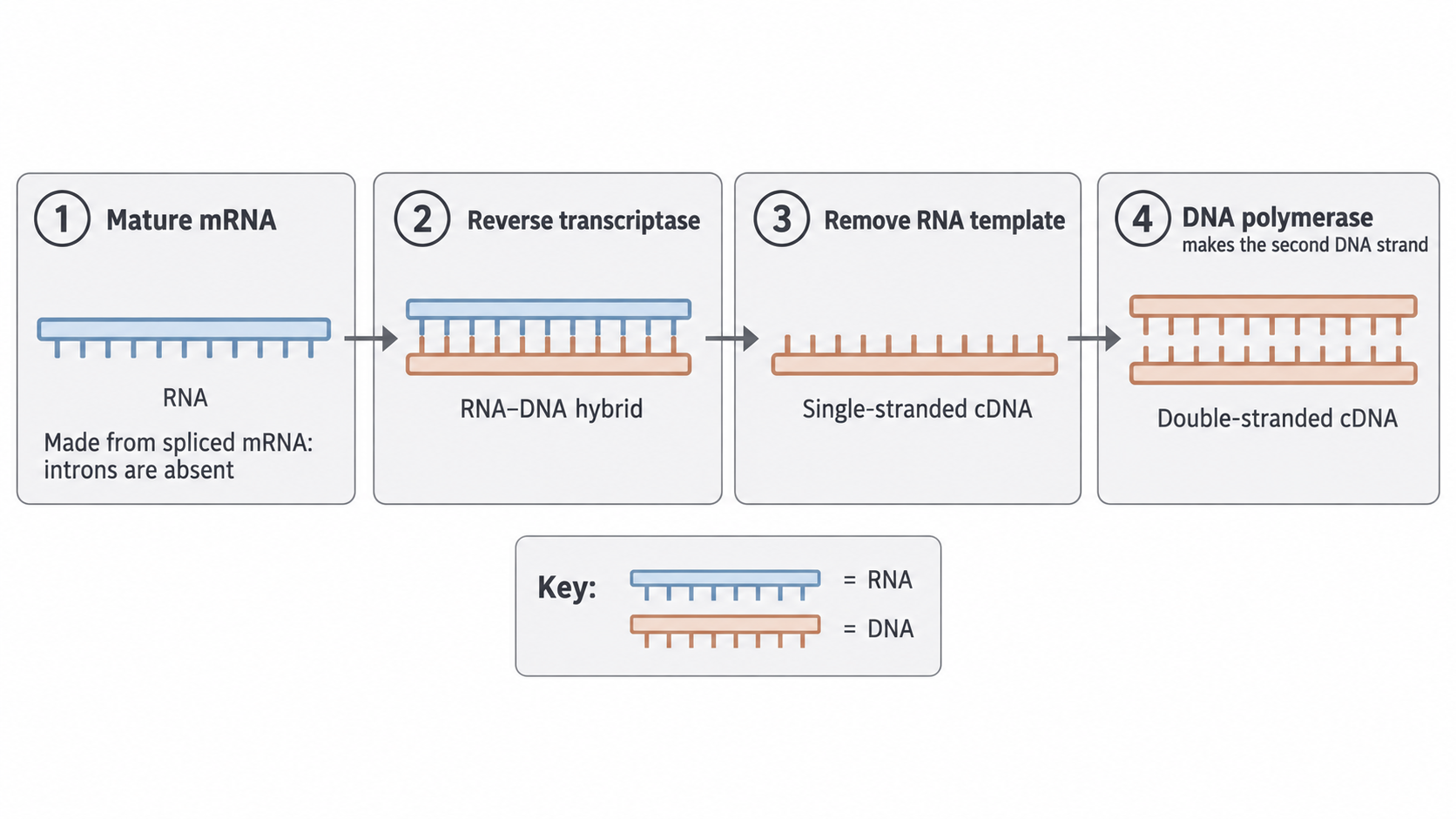
- Extract mature mRNA from a cell type that expresses the gene abundantly, such as the beta cells of the islets of Langerhans.
- Make the first DNA strand. Reverse transcriptase joins free DNA nucleotides that are complementary to the mRNA, giving an RNA–DNA hybrid. The new strand is complementary DNA (cDNA).
- Remove the RNA template, leaving single-stranded cDNA.
- Make the second strand. DNA polymerase adds nucleotides complementary to the cDNA template, producing a double-stranded DNA copy of the gene.
Complementary DNA (cDNA)
DNA synthesised from an RNA template. Reverse transcriptase first makes one complementary DNA strand; removal of the RNA and synthesis of a second DNA strand produce double-stranded cDNA. cDNA made from mature, spliced mRNA lacks the introns removed during processing.
Mature mRNA has already been spliced, so its cDNA has no introns. This matters because prokaryotic cells lack the machinery to remove introns: a bacterium can use the intron-free coding sequence when suitable control sequences are supplied. Eukaryotic proteins that require particular processing or modifications may still need a different host.
Restriction endonucleases
The second route cuts a fragment containing the gene directly out of the organism's DNA.
Restriction Endonuclease
An enzyme, produced naturally by bacteria as a defence against viral DNA, that cuts double-stranded DNA at a specific base sequence known as a recognition site.
Each enzyme's active site is complementary to a specific short base sequence, so different enzymes cut at different sites and cuts are not random.
At its site, the enzyme breaks phosphodiester bonds in both DNA strands. To remove a gene, use an enzyme whose recognition sites lie either side of it. The fragment keeps any introns the gene has.
The way the enzyme cuts decides what you can do with the fragment:
- Blunt ends: the enzyme cuts straight across both strands at the same position. HpaI recognises GTTAAC and cuts between the T and A on both strands.
- Sticky ends: the enzyme makes a staggered cut, leaving short single-stranded overhangs of unpaired bases. HindIII recognises AAGCTT and cuts between the two A bases on each strand. AAGCTT reads the same 5′ to 3′ on both strands, so the two overhangs are complementary.
Sticky Ends
Short, single-stranded overhangs of unpaired nucleotide bases produced when a restriction endonuclease makes a staggered cut through double-stranded DNA. Sticky ends from the same enzyme are complementary and can base-pair with each other.
An enzyme recognises the same short sequence wherever it occurs, so it leaves matching sticky ends on DNA from any organism. This is what allows fragments from different species to be joined, the basis of recombinant DNA.
Compatible sticky ends make it easier to align an insert with a vector. Blunt-ended DNA can also be joined, but it lacks this initial pairing.
The gene machine
The third route needs no DNA or RNA template, because the gene is built from its sequence.
- If the protein's amino acid sequence is known, scientists use the genetic code to design a possible coding DNA sequence. The code is degenerate, so more than one DNA sequence may code for the protein: the sequence is designed, not uniquely read off.
- A computer designs a series of short, overlapping single-stranded sequences called oligonucleotides.
- An automated synthesiser assembles each oligonucleotide one nucleotide at a time.
- The oligonucleotides are joined to form the gene, the complementary strand is synthesised, and the finished gene can be copied by PCR (next section).
A chosen sequence can be produced rapidly and with high accuracy, designed without introns and with codons suitable for the host. Expression still requires compatible regulatory sequences: synthesising the DNA alone does not produce protein.
| Route | Starts from | Key tool | Introns |
|---|---|---|---|
| cDNA | Mature mRNA | Reverse transcriptase | Removed by splicing |
| Restriction | Extracted DNA | Restriction endonuclease | Kept, if the gene has any |
| Gene machine | Known sequence | Automated synthesiser | Left out of the design |
Amplifying DNA fragments
One fragment is far too little DNA to work with, so it is copied, either in a tube or inside living cells.
The tube method, PCR, is in vitro: outside living cells. The other method is in vivo: the culture of host cells that have taken up the DNA, called transformed cells (see transformation, in the next section).
The polymerase chain reaction (PCR)
Polymerase Chain Reaction (PCR)
An in vitro technique that produces millions of copies of a specific DNA fragment through repeated cycles of heating and cooling, using a thermostable DNA polymerase.
PCR is carried out in a thermocycler, a programmable machine that precisely controls temperature. The reaction mixture contains:
- the target DNA fragment
- free DNA nucleotides
- two short primers, single-stranded sequences complementary to the ends of the target fragment
- a heat-stable DNA polymerase called Taq polymerase, from the hot-spring bacterium Thermus aquaticus
The principle is that each strand made in one cycle becomes a template in the next. Each cycle has three temperature-controlled steps, shown left to right in the diagram.
[DIAGRAM: asset_name: 8.4.1 - Recombinant DNA technology - Diagram 2; asset_slug: 8.4.1 - Recombinant DNA technology - Diagram 2; recommended_method: retained_png; caption: One PCR cycle separates strands, binds primers and extends them, repeated ideal cycles double the DNA copies.; description: One PCR cycle separates strands, binds primers and extends them; repeated ideal cycles double the DNA copies.]
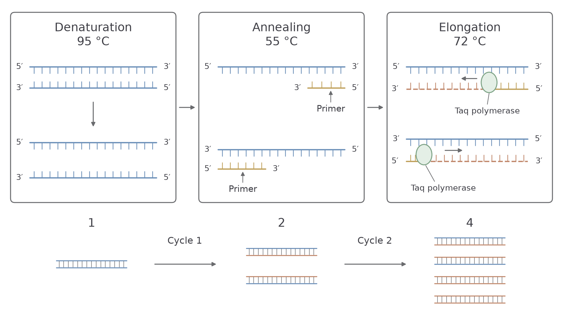
- Denaturation (95 °C). The high temperature breaks the hydrogen bonds between complementary base pairs, separating the DNA into two single strands.
- Annealing (often around 50–65 °C). Cooling lets the primers bind (anneal) to complementary sequences at each end of the target. Primers are in excess, which favours this binding, although strands can also re-anneal. DNA polymerase can only add nucleotides to the end of an existing strand, so primers provide the starting point. Synthesis begins only where a primer has bound, so only the DNA lying between the two primers is amplified.
- Elongation (about 72 °C for Taq). This is close to Taq's optimum temperature. Taq extends each primer by adding complementary nucleotides along the template strand, giving two double-stranded copies of the target.
Taq is thermostable, so it survives the 95 °C steps and the same enzyme can work through later cycles. Annealing temperature depends on the primers; the diagram's 55 °C is one possible value.
The lower row of the diagram shows the result: in the idealised model each cycle doubles the number of target molecules, so one becomes two, then four.
Number of DNA molecules after PCR
is the starting number of target molecules and is the number of complete cycles.
Use this model when a question states perfect doubling. Actual reactions become less efficient as reagents are used up.
Worked example: counting PCR products
A PCR starts with 2 target DNA molecules. Assuming perfect doubling, calculate the number after 10 complete cycles. Give your answer in standard form to 3 significant figures.
Plan. Substitute into the formula, and round only the final result.
Substitute.
Standard form.
Answer. molecules to 3 significant figures.
Check. Ten doublings multiply by about 1000, so the answer should be close to 2000.
In vivo amplification, compared with PCR
The alternative is to clone the DNA inside living cells. The fragment is inserted into a vector, a carrier such as a plasmid (the next section shows how). The vector is introduced into bacterial host cells, which are cultured.
As these transformed cells grow and divide, the vector replicates, and the inserted DNA with it, giving many copies of the gene. PCR is often faster for obtaining DNA. The advantage of culture is that it maintains a clone and can make the gene's protein product.
Neither method is automatically error-free: polymerase choice, culture conditions and sequence checking all matter. Contamination troubles PCR when the contaminating DNA contains the target region recognised by the primers. PCR does not amplify every contaminating sequence.
| PCR (in vitro) | Cell culture (in vivo) | |
|---|---|---|
| Copies | A selected DNA region | The whole recombinant vector |
| Happens | In a tube, with Taq | Inside living host cells |
| Speed | Often faster for DNA | Slower |
| Makes protein? | No | Can transcribe and translate the gene |
Cover the table, then explain from memory how the two methods differ.
Inserting DNA and transforming hosts
A gene needs control regions and a carrier before a host cell will copy it and express it.
For in vivo cloning, the carrier is a vector. The most commonly used vectors are plasmids: small, circular DNA molecules found naturally in bacteria, separate from the main bacterial chromosome.
Preparing the fragment with control regions
A coding sequence alone is not enough, because transcription needs a start and a stop. A promoter is not transcribed, so a cDNA copy of mature mRNA has none. Suitable regulatory sequences must be provided, either added to the fragment or already positioned in an expression vector:
- A promoter region is a DNA sequence upstream of the gene. RNA polymerase, with transcription factors in eukaryotes, binds here to initiate transcription.
- A terminator region is a sequence downstream of the gene. It causes RNA polymerase to stop transcription and detach.
The promoter must be recognised by the host machinery and orientated correctly relative to the coding sequence. The terminator ends transcription; it is not the same as a stop codon, which ends translation.
Vector
A carrier molecule, such as a plasmid, used to transfer a fragment of DNA into a host cell. Plasmid vectors have an origin of replication, allowing their DNA to be copied in a suitable host; expression vectors also supply compatible control sequences.
Joining the fragment to the plasmid
Cut a plasmid and a fragment with the same restriction endonuclease and their sticky ends are complementary, so they can pair. A second enzyme then makes the join permanent.
DNA Ligase
An enzyme that joins two fragments of DNA by catalysing the formation of phosphodiester bonds between the sugar–phosphate backbones, sealing nicks in the double helix.
Follow panels 1 to 3 of the diagram. The strip beneath them shows the order of the parts needed for expression: promoter, coding sequence, terminator.
[DIAGRAM: asset_name: 3.8.4.1-recombinant-v2; asset_slug: 3.8.4.1-recombinant-v2; recommended_method: retained_png; caption: Compatible sticky ends base-pair before ligase seals the DNA backbones, expression needs suitable control sequences.; description: Compatible sticky ends base-pair before ligase seals the DNA backbones; expression needs suitable control sequences.]
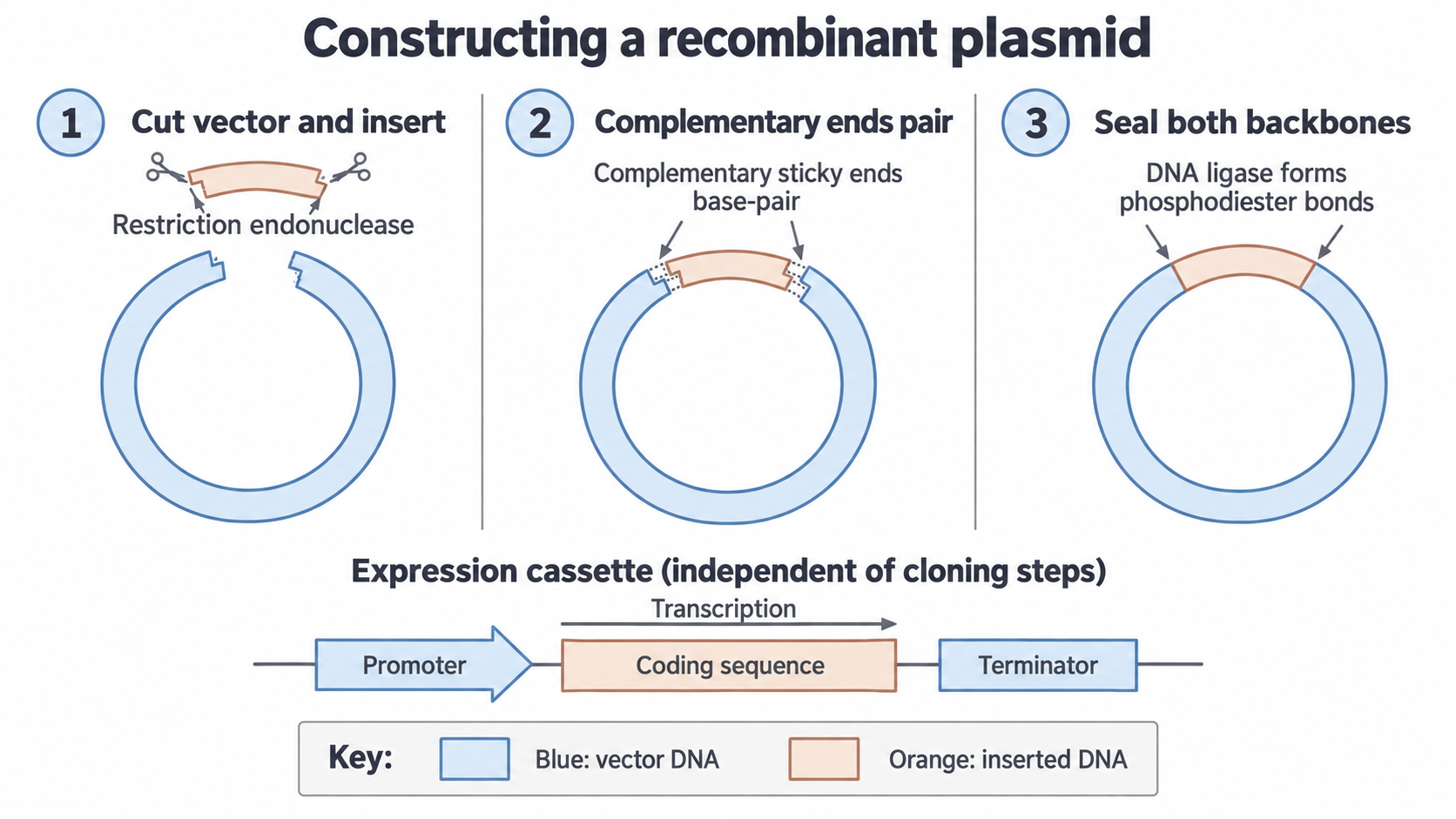
- The restriction endonuclease cuts the plasmid at a suitable recognition site; in some designs the site lies within a marker gene.
- The DNA fragment, with its matching sticky ends, is mixed with the opened plasmids.
- Complementary sticky ends base-pair through hydrogen bonding.
- DNA ligase catalyses the formation of phosphodiester bonds, sealing the sugar–phosphate backbone on both strands and permanently joining the fragment into the plasmid.
The result is a recombinant plasmid: a plasmid containing DNA from two different sources.
Watch out
Weaker exam answers say DNA ligase cuts the plasmid or creates the sticky ends. It does neither: the restriction endonuclease cuts and leaves sticky ends, and ligase forms phosphodiester bonds. Saying that ligase joins base pairs can lose the mark, because those hydrogen bonds form without it.
Transformation
The recombinant plasmids now have to get into the host cells.
Transformation
The process by which a host cell takes up foreign DNA, such as a recombinant plasmid, from its surroundings.
The plasmids are mixed with bacterial host cells in a medium containing calcium ions (Ca²⁺). A brief heat shock follows, a rapid temperature shift from around 0 °C to approximately 42 °C. Together these increase the permeability of the bacterial cell-surface membrane, so plasmids can pass into the cytoplasm.
Only a proportion of cells take up a plasmid, depending on the method and conditions. Some of those take up a plasmid with no fragment, because a cut plasmid's two sticky ends are complementary to each other, so it can close back up. Identifying the right cells is therefore the next step.
Continue this lesson.
You’re previewing NovaLearn without an account. Create a free account to read the rest.
Create free account