3.2.3 - Transport across cell membranes
A cell stays alive because its membrane lets some substances through and holds others back. You’ll see how the fluid-mosaic structure does this, the five routes across, and how specialised cells speed them up.
Membrane structure
Two layers of lipid and a scatter of proteins decide what enters and leaves a cell.
Cell-surface membranes and the membranes around eukaryotic organelles share the same basic structure, described by the fluid-mosaic model. The proteins present vary with each membrane’s function.
Fluid-Mosaic Model
A model in which phospholipids form a fluid bilayer and proteins are scattered through it like a mosaic. Individual phospholipids can move sideways within the layer.
The arrangement of components
Start with the bilayer in the diagram. Phosphate heads are hydrophilic and fatty acid tails are hydrophobic (3.1.3), so the heads face the watery environments on either side and the tails face inwards. Proteins lie within or across the bilayer, and cholesterol fits between phospholipids rather than forming a separate layer.
[DIAGRAM: asset_name: 2.3 - Transport across cell membranes - Diagram 1; asset_slug: 2.3 - Transport across cell membranes - Diagram 1; recommended_method: retained_png; caption: A labelled fluid-mosaic membrane showing phospholipid bilayer, channel protein, carrier protein, glycoprotein, glycolipid and cholesterol.; description: A labelled fluid-mosaic membrane showing phospholipid bilayer, channel protein, carrier protein, glycoprotein, glycolipid and cholesterol.]
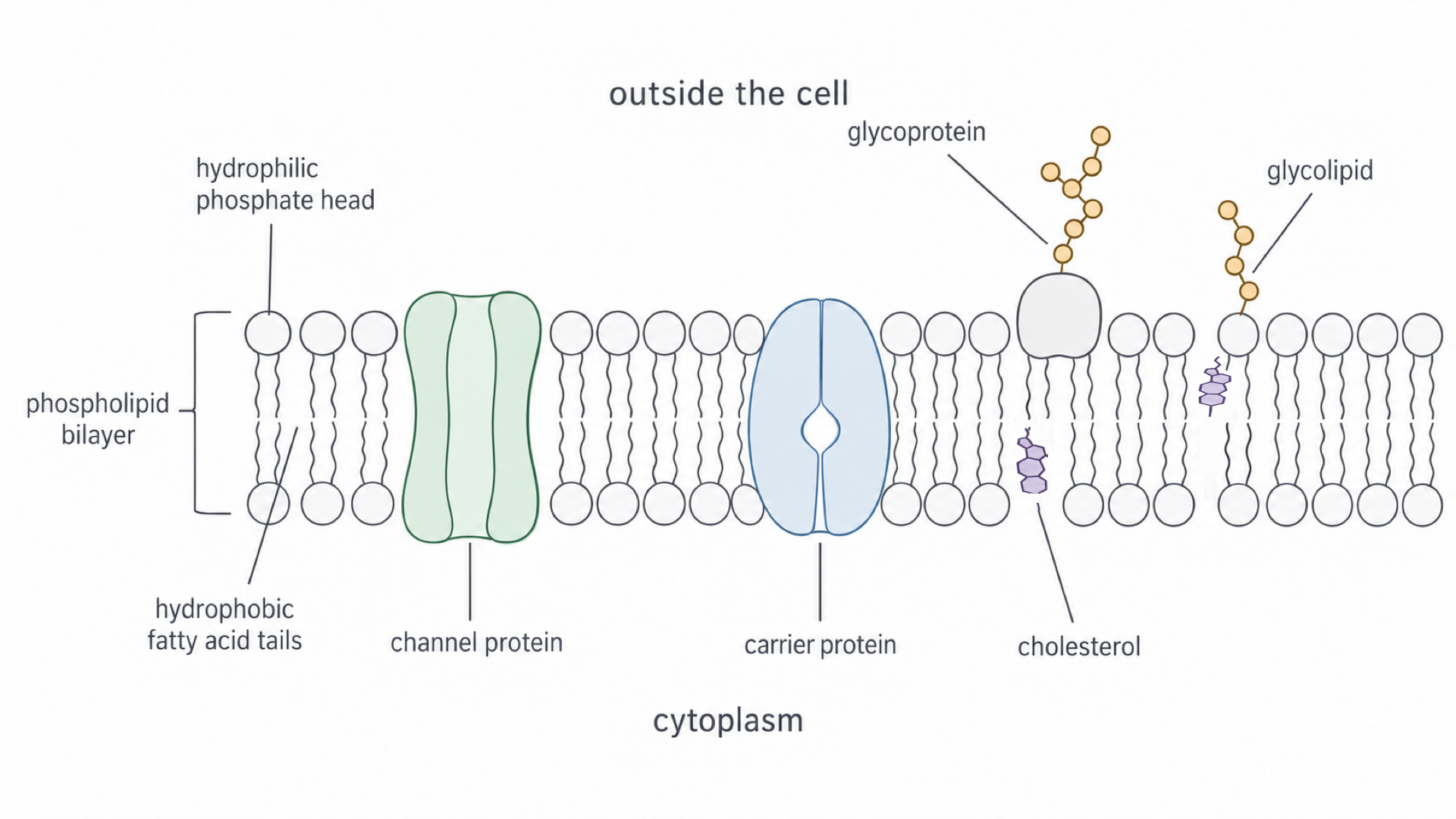
| Component | Where it sits | Main role |
|---|---|---|
| Phospholipids | Two layers, tails inwards | Form the bilayer, a barrier to many water-soluble substances |
| Channel proteins | Across the bilayer | Form hydrophilic pores for particular ions or water molecules |
| Carrier proteins | Across the bilayer | Bind a specific substance and change shape to move it across |
| Glycoproteins | In the bilayer: a protein with a carbohydrate chain attached | Cell recognition and receptors |
| Glycolipids | In the bilayer: a lipid with a carbohydrate chain attached | Recognition and membrane stability |
| Cholesterol | Between phospholipids | Makes the membrane more stable and less permeable to water and ions |
The carbohydrate chains of glycoproteins and glycolipids project from the surface facing away from the cytoplasm.
What moves and what restricts it
The membrane is fluid because phospholipids move sideways (laterally) within their layer. Many proteins and other lipids drift sideways too, although some proteins are anchored.
Cholesterol may also be present, and it restricts the movement of the other molecules in the membrane. So the membrane becomes less fluid and more stable, and less water and fewer ions leak through it.
Diffusion and facilitated diffusion
Oxygen crosses a membrane with no help, but glucose barely crosses at all without a protein.
Diffusion is the net movement of particles from a region of higher concentration to a region of lower concentration. It is passive: particles move using their own kinetic energy, so the cell does not have to supply any.
The simplest case is simple diffusion directly through the phospholipid bilayer. The hydrophobic middle of the bilayer limits what can cross this way:
- Small non-polar molecules, such as oxygen and carbon dioxide, pass through quite easily.
- Charged ions and polar molecules, such as glucose and amino acids, do not dissolve in the hydrophobic core, so they barely cross.
- Large molecules do not slip easily between the phospholipids.
Channel and carrier proteins
Substances the bilayer blocks can cross through proteins instead.
Facilitated Diffusion
The passive movement of a substance down its concentration gradient through a membrane protein.
Two kinds of protein do this, each specific to particular substances:
- Channel proteins provide a hydrophilic pore through the membrane. This route is especially important for ions.
- Carrier proteins bind a specific molecule, such as glucose, then change shape and release it on the other side.
The protein provides a route, not energy. Like simple diffusion, facilitated diffusion moves a substance down its concentration gradient and uses no ATP.
Exam tip
Write “channel protein” or “carrier protein”. “Transport protein” or just “protein” can lose marks.
What sets the rate
Particles cross in both directions, but many more cross from the high-concentration side than cross back. So a steeper concentration gradient gives more net movement per second. The rate also increases with:
- a larger membrane surface area, because more particles can cross at once
- a shorter diffusion distance, because particles have less far to travel
- more suitable channel or carrier proteins, because there are more routes across
Osmosis and water potential
Put a red blood cell in pure water and it bursts. Put a plant cell in the same water and it simply firms up.
Osmosis is a special case of diffusion because it involves only water.
Osmosis
The net movement of water molecules from a region of higher water potential to a region of lower water potential through a partially permeable membrane.
A partially permeable membrane lets water cross but holds back many solutes. Which way the water moves depends on water potential.
Water Potential
A measure of the tendency of water molecules to move from one place to another.
Water potential is measured in kPa. Pure water at atmospheric pressure has a water potential of 0 kPa. Adding solute lowers it: solute particles attract water molecules, which leaves fewer free to move, so the water potentials of solutions are negative.
The less negative value is the higher water potential. So water moves from a solution at −100 kPa into a cell initially at −400 kPa.
Watch out
Don’t explain osmosis with “water concentration” or “high to low concentration”. This is ambiguous, because a solution with a high solute concentration has a low water potential, and it can lose marks. Write from a higher to a lower water potential.
How water crosses and how fast
Water molecules are polar, but they are small enough for some to pass through the bilayer. Water channels called aquaporins give a much faster route. Neither route requires ATP.
A larger water potential difference produces faster net movement through an otherwise unchanged membrane. As water moves, the difference shrinks, so net movement slows. At equal water potentials molecules still cross in both directions, but at equal overall rates.
Why animal and plant cells respond differently
The outcome differs because a plant cell has a cell wall.
An animal cell has no wall to resist expansion, so in a much higher water potential it swells and may burst. As a plant cell takes up water, its contents press against the wall. This pressure raises the cell’s water potential until it equals the external value, stopping net uptake.
A cell pressed firmly against its wall like this is turgid; a cell with no pressure against its wall is flaccid. So a turgid cell can be at water-potential equilibrium: equal water potentials do not automatically mean a flaccid cell.
Continue this lesson.
You’re previewing NovaLearn without an account. Create a free account to read the rest.
Create free account