3.1.5.1 - Structure of DNA and RNA
DNA and RNA are the information-carrying molecules of every living cell: DNA holds genetic information and RNA transfers it to the ribosomes. Build both from nucleotides, tell apart the bonds along and between strands, and use base pairing to work out sequences and percentages.
Nucleotides
Deoxyribonucleic acid (DNA) and ribonucleic acid (RNA) are both polymers of one kind of unit: the nucleotide.
Nucleotide
A nucleotide is the monomer from which a nucleic acid is built. It is formed from a pentose sugar, a nitrogen-containing organic base and a phosphate group.
In the diagram, the pentagon is the pentose sugar, a sugar with five carbon atoms. The phosphate group and the nitrogen-containing organic base both attach to it.
[DIAGRAM: asset_name: nucleotide-structure; asset_slug: nucleotide-structure; recommended_method: retained_png; caption: A single nucleotide: a phosphate group and a nitrogen-containing organic base, each attached to a pentose sugar.; description: A single nucleotide: a phosphate group and a nitrogen-containing organic base, each attached to a pentose sugar.]
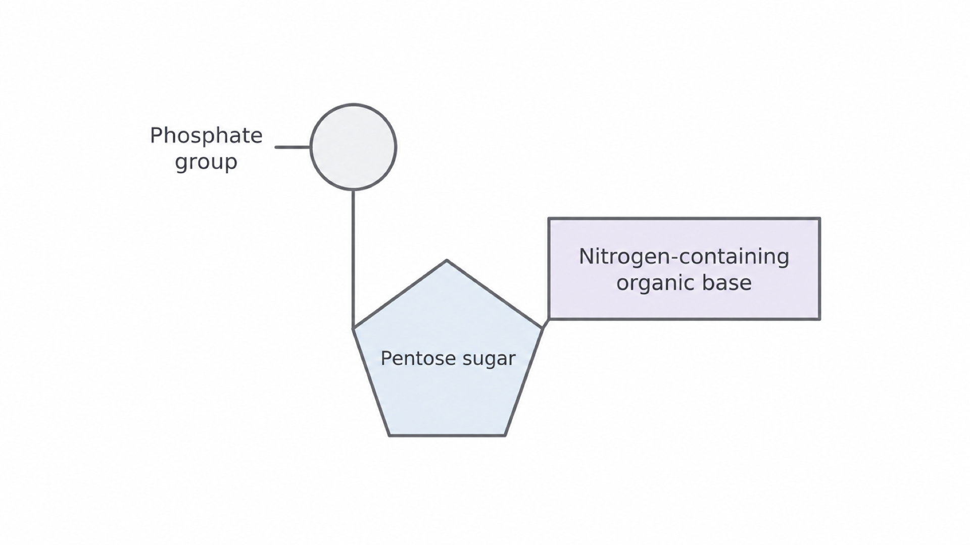
A single nucleotide contains one of the four possible bases, not all four.
| Component | DNA nucleotide | RNA nucleotide |
|---|---|---|
| Pentose sugar | Deoxyribose | Ribose |
| Phosphate | Phosphate group | Phosphate group |
| One possible base | Adenine (A), thymine (T), cytosine (C) or guanine (G) | Adenine (A), uracil (U), cytosine (C) or guanine (G) |
Exam tip
Pair each difference: deoxyribose in DNA and ribose in RNA, thymine in DNA and uracil in RNA. “Double-stranded” describes a whole DNA molecule, not a nucleotide.
Cover the table, then list the components of each nucleotide from memory.
From nucleotides to polynucleotides
Nucleotides join by condensation, the reaction that also builds starch and proteins.
Phosphodiester bond
A phosphodiester bond is the strong covalent bond formed by a condensation reaction between two nucleotides. It links the phosphate group of one nucleotide to the pentose sugar of the next.
Each of these condensation reactions releases one molecule of water and forms one bond. So a chain of 5 nucleotides has 4 phosphodiester bonds, and making it releases 4 water molecules.
The phosphate links the sugars of neighbouring nucleotides, producing a repeating sugar–phosphate backbone with a base projecting from each sugar. The result is a polynucleotide, meaning a chain of many nucleotides.
Where the information sits
The backbone repeats, but the base can vary from one nucleotide to the next, so the information lies in the order of bases. Two chains can contain the same numbers of each base but carry different sequences, just as “listen” and “silent” use the same letters to make different words.
RNA structure and roles
In all living cells, DNA holds genetic information and RNA transfers it to the ribosomes, where proteins are made.
An RNA molecule is a relatively short, single polynucleotide chain compared with cellular DNA. Single-stranded does not mean straight: a chain can fold, and regions of that same chain can pair with each other.
In a eukaryotic cell the nuclear DNA stays in the nucleus but ribosomes are in the cytoplasm, so messenger RNA (mRNA) leaves through nuclear pores to reach them, carrying a copy of the information. At the ribosome it directs protein synthesis.
Ribosomes are made of ribosomal RNA (rRNA) and proteins. They are not made of protein alone. Transfer RNA (tRNA) is a third type, which carries amino acids during protein synthesis.
| Type of RNA | Role |
|---|---|
| mRNA | Carries a copy of genetic information from DNA to a ribosome |
| rRNA | Forms ribosomes, together with proteins |
| tRNA | Carries amino acids during protein synthesis |
“RNA” alone does not say which of these you mean, so name the type.
The structure of DNA
Picture a twisted ladder.
DNA has that shape: two polynucleotide strands wound into a double helix. The sugar–phosphate backbones are the sides, on the outside, and pairs of bases are the rungs.
Hydrogen bonds between the bases hold the two strands together. The pairing is specific: A pairs with T, and G pairs with C, because the shapes and bonding groups of the bases line up only in those pairs.
Bases joined like this are complementary base pairs, so the sequence on one strand determines the sequence on the other. Directly opposite A G T C lies T C A G. The complementary strand is not an identical copy of the first strand.
In the diagram, follow one strand down: phosphate, deoxyribose, phosphate. The bracket marks one phosphodiester linkage. The dots are hydrogen bonds: two in each A–T pair and three in each G–C pair. On the right, RNA has one chain, with ribose and U in place of T.
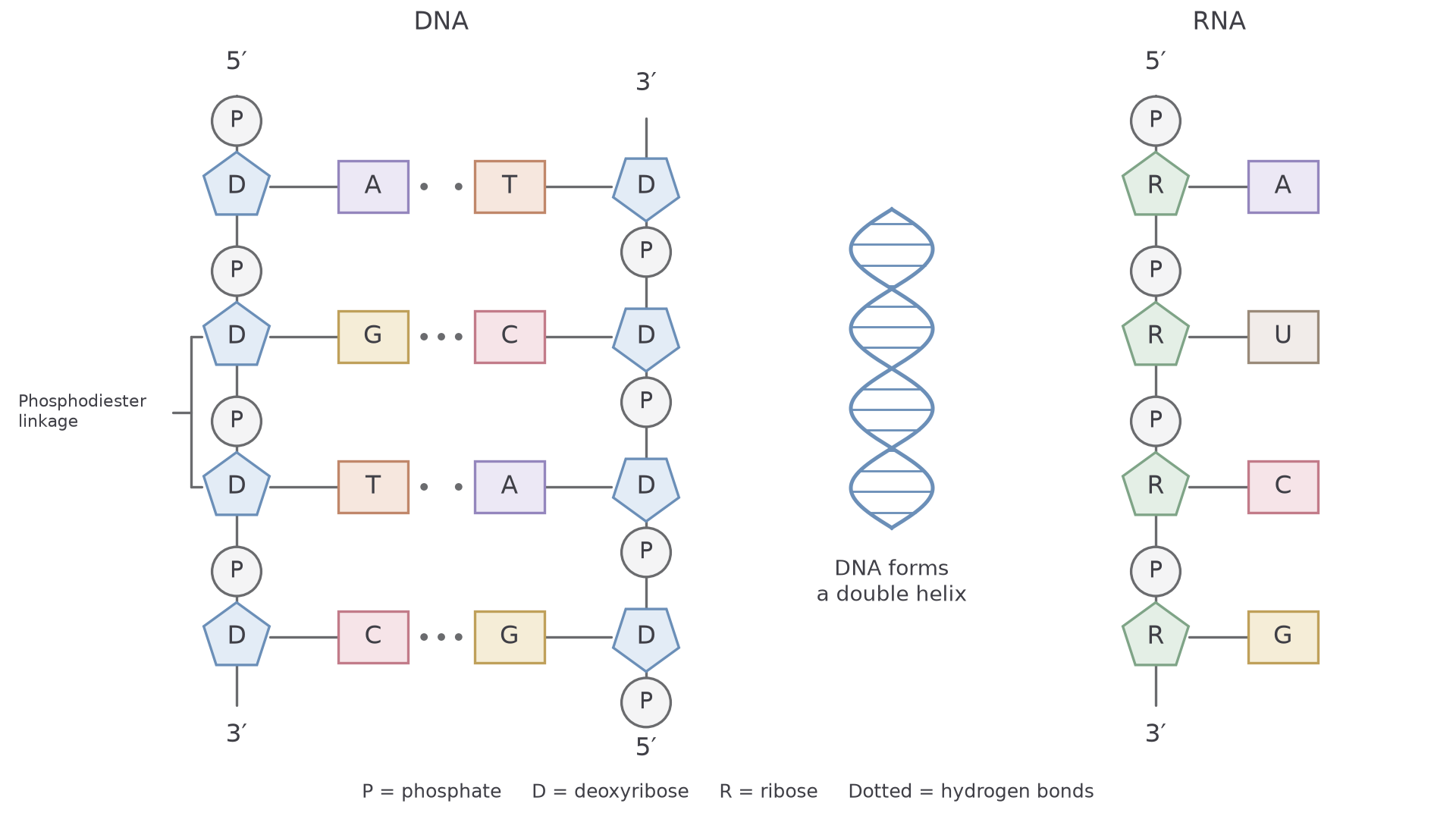
Every backbone link runs the same way, so each strand has a direction, named by its 5′ and 3′ ends. 5′ and 3′ are numbered sugar carbons; they do not count bases. In the diagram one strand runs 5′ to 3′ and the other 3′ to 5′: they are antiparallel.
Watch out
Phosphodiester bonds join adjacent nucleotides along one strand. Hydrogen bonds join complementary bases between strands. Don’t swap them.
Using base frequencies
Every A is paired with one T and every G with one C. So in a complete double-stranded DNA molecule, the numbers of A and T are equal, as are those of G and C. The four percentages add to 100%.
Worked example: from one percentage to a base count
Question. A DNA molecule has 600 base pairs, and 28% of all its bases are adenine. Find the number of cytosine bases.
Plan. Find the percentage of C, then apply it to bases, not base pairs.
- A pairs with T, so T is also 28%, leaving 44% for G plus C.
- G and C are equal, so .
- Each pair holds two bases, so 600 base pairs means 1200 bases, and 22% of 1200 is 264.
Answer. 264 cytosine bases (and 264 guanine).
Check. A and T are each 336, and 336 + 336 + 264 + 264 = 1200.
Frequencies on a single strand
These equalities need not hold within one strand, because each base’s partner is on the other strand. If one strand is 35% A, its complementary strand is 35% T, but that fact alone says nothing about the T or C in the first strand.
To complete a strand, work across the pair: each base’s percentage in one strand equals its partner’s percentage in the other.
| Base | Strand 1 (%) | Strand 2 (%) | Both strands (%) |
|---|---|---|---|
| A | 35 | 20 | 27.5 |
| T | 20 | 35 | 27.5 |
| G | 30 | 15 | 22.5 |
| C | 15 | 30 | 22.5 |
Across both strands, A and T are each , and G and C are each . RNA is a single chain, so its A need not equal its U.
Why DNA is a stable information store
DNA keeps its base sequence intact, yet its strands can separate for copying. Its two bond types explain both.
Strong covalent phosphodiester bonds in each backbone preserve the order of nucleotides. Many hydrogen bonds collectively hold the strands together. The bases lie inside the molecule, while the backbones form its exterior, so the backbones help to shield the information-carrying bases from chemical damage.
An individual hydrogen bond is much weaker than a covalent backbone bond. Enzymes can therefore break the hydrogen bonds and separate the strands locally, which exposes their bases without breaking either backbone. Complementary pairing then allows each strand to act as a template for a new partner.
Long DNA molecules carry information in enormous numbers of possible base sequences. Length gives capacity; the order of bases specifies the information. So neither “DNA contains four bases” nor “DNA is long” explains how two DNA molecules can hold different messages.
Why scientists once doubted DNA
For decades, many scientists expected genes to be made of protein, not DNA.
Proteins contain twenty different amino acids with varied chemical properties, while DNA uses only four base types.
An influential early model made DNA look simpler still, picturing it as one unit of four nucleotides repeated unchanged. This relative simplicity led many scientists to doubt that DNA carried the genetic code.
The mistake was to confuse a small number of building blocks with a small number of possible arrangements. Even a four-position sequence has possible arrangements if each position can hold any of four bases, and each extra position multiplies the total by four.
Equal overall base percentages also do not imply equal sequences: AATT and ATAT contain the same base numbers but different orders.
This reasoning shows only that DNA could have sufficient information capacity. Proving it was the hereditary material took experiments. In 1944 Avery, MacLeod and McCarty showed that DNA caused a heritable change in bacteria: removing protein left the effect intact, but enzymes that digest DNA destroyed it.