3.6.1.2 - Receptors
Receptors turn a specific stimulus into an electrical signal. See how in the Pacinian corpuscle, how the pigments and wiring of rods and cones shape vision, and how to test skin sensitivity.
The Pacinian corpuscle as a receptor
Press firmly on your skin and a layered capsule deep below turns that push into an electrical signal.
Receptors are specific: each type responds only to a particular kind of stimulus. Converting the stimulus's energy into an electrical change is called transduction.
Receptor
A cell or group of cells that detects a stimulus and converts the energy of that stimulus into an electrical signal.
The Pacinian corpuscle is a mechanoreceptor, a receptor for mechanical stimuli. It lies deep in the skin and also in some joints, tendons and ligaments, and it responds to mechanical pressure and vibration.
Pacinian corpuscle
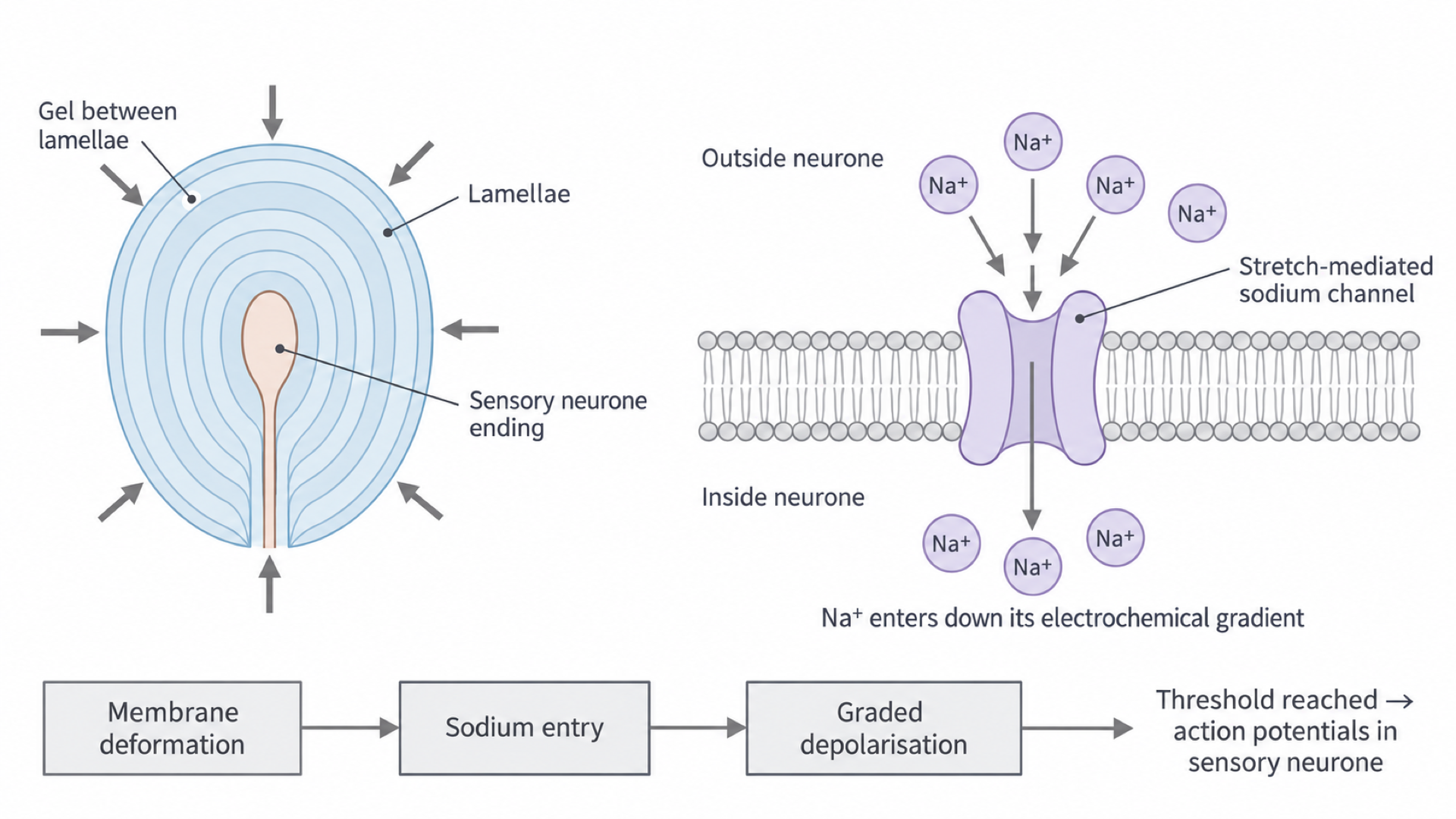
A pressure receptor made of the ending of a sensory neurone surrounded by concentric layers of connective tissue called lamellae, with gel between the layers.
In cross-section it looks like an onion, as the next section's diagram shows.
Why it responds to pressure
The membrane of the sensory neurone contains stretch-mediated sodium ion channels, which open when the membrane around them is stretched or deformed. A stimulus that does not deform the membrane, such as light or a small temperature change, leaves them closed. That is why this receptor responds to pressure and not to other stimuli.
From pressure to a generator potential
Follow a squeeze from the outer layers of the corpuscle to the first nerve impulse.
Nerve impulses are taught fully in 3.6.2.1, but you need four terms here:
- Membrane potential: the voltage across a neurone's membrane.
- Resting potential: the membrane potential of an unstimulated neurone, with the inside negative compared with the outside.
- Action potential: a brief, large change in membrane potential that travels along the neurone as a nerve impulse.
- Threshold: the membrane potential that must be reached before an action potential starts.
Generator potential
A change in membrane potential produced in a receptor when a stimulus opens ion channels. If it reaches threshold, an action potential is triggered in the sensory neurone.
How pressure opens sodium channels
With no pressure, the membrane is not deformed, the stretch-mediated sodium ion channels stay closed, and the neurone remains at its resting potential. Pressure sets off a chain:
- Pressure compresses the lamellae, which deforms the membrane of the sensory neurone.
- The deformation opens the stretch-mediated sodium ion channels.
- Sodium ions diffuse into the neurone down their electrochemical gradient: they are more concentrated outside, and the negative inside attracts their positive charge.
- This entry of positive charge makes the inside less negative, so the membrane is depolarised. This depolarisation is the generator potential.
- If the generator potential reaches threshold, an action potential is produced, and the impulse travels along the sensory neurone to the central nervous system.
In the diagram, the left drawing shows step 1, the right steps 2 and 3, and the bottom strip the whole chain.
Graded potentials, all-or-nothing impulses
A generator potential is graded: stronger pressure deforms the membrane more, so more channels open, more sodium ions enter, and the local depolarisation is larger. Below threshold, there may be a generator potential without any action potential.
For example, if resting potential is −70 mV and threshold is −55 mV, a generator potential of 8 mV brings the membrane to −62 mV, below threshold. A 17 mV depolarisation reaches −53 mV, beyond threshold. These values are illustrative.
Action potentials differ. Their amplitude is all-or-nothing: each one in that neurone is the same size however strong the stimulus. Stronger stimulation can increase their frequency instead, so do not draw a larger action potential to represent greater pressure.
Why sustained pressure fades
Pacinian corpuscles respond especially to changes in pressure and vibration. During sustained pressure the layers can redistribute the force, so deformation of the sensory ending falls and its response diminishes.
This is receptor adaptation; it does not mean the sodium supply has run out. It also differs from habituation, a decrease in response after repeated harmless stimulation involving processing in the nervous system.
Photoreceptors and the retina
Light must pass through layers of nerve cells before it reaches the rods and cones that detect it.
Where light lands in the eye
Start at the left of the diagram. Light enters through the cornea, then passes through the pupil (an opening) and the lens. The cornea and lens focus it on the retina, the light-sensitive layer at the back of the eye.
[DIAGRAM: asset_name: eye-structure; asset_slug: eye-structure; recommended_method: retained_png; caption: Section through the human eye showing light focused on the fovea, with cone cells concentrated at the fovea, rod cells in the peripheral retina, and the blind spot where the optic nerve leaves.; description: Section through the human eye showing light focused on the fovea, with cone cells concentrated at the fovea, rod cells in the peripheral retina, and the blind spot where the optic nerve leaves.]
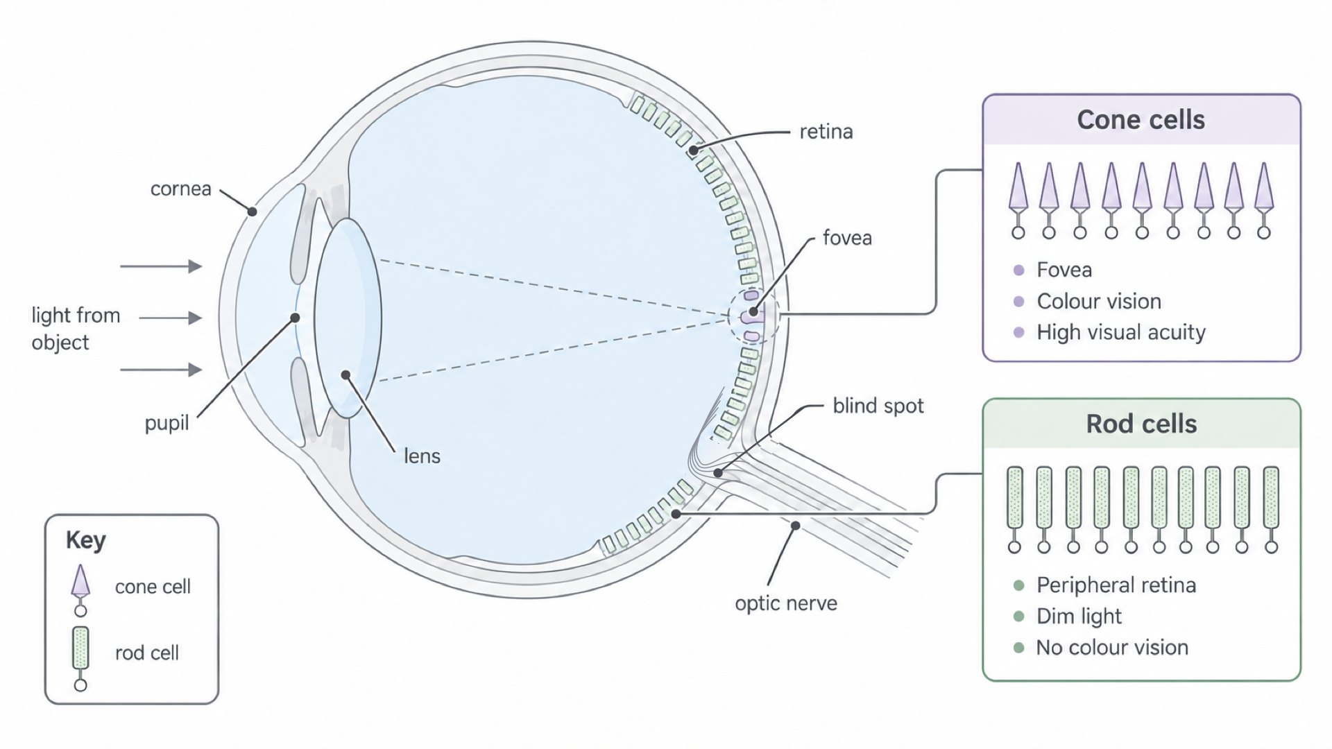
The dashed lines end at the fovea: whatever you look at directly is focused here. This small central region has a very high cone density. Its centre has no rods, and the inner retinal layers are pushed aside, so light reaches the cones with less scattering.
Light from anything to the side of your gaze lands on the peripheral retina, where rods are numerous. The panels show where each cell type is concentrated: cones also occur, more sparsely, outside the fovea.
The blind spot is where optic-nerve axons leave the eye. It has no photoreceptors, so no light can be detected at that point.
From photoreceptor to optic nerve
Rods and cones are photoreceptors: their light-sensitive pigments absorb light, and this transduces it into a graded electrical response. In most of the retina, incoming light passes through the ganglion-cell and bipolar-cell layers first. Information then passes in the opposite overall direction:
Photoreceptor → bipolar cell → ganglion cell → optic nerve
The axons of the ganglion cells form the optic nerve, which carries action potentials to the brain. Rods and cones themselves produce only graded responses.
In the next diagram light travels up the page and signals travel down it.
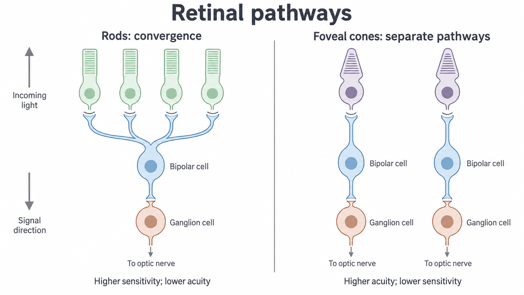
This simplified diagram is not to scale. Count the photoreceptors sharing an output: four rods share one ganglion cell, while each foveal cone has its own.
That count shapes sensitivity, the ability to detect a weak stimulus, and visual acuity, the ability to distinguish two close points as separate.
Continue this lesson.
You’re previewing NovaLearn without an account. Create a free account to read the rest.
Create free account