3.2.2 - Cell division
Every cell in your body came from an earlier cell. Follow the DNA through the cell cycle, mitosis and cytokinesis, then see what uncontrolled division causes, how cancer drugs slow it, how prokaryotes divide by binary fission and why viruses do not divide at all.
The cell cycle
A cell copies its DNA before it divides, so the time between divisions is anything but rest.
Every new cell comes from a pre-existing cell, but not all cells retain the ability to divide. Many mature specialised cells leave the cycle, whereas stem cells and meristematic cells (found in plant root and shoot tips) can divide. Cells that do divide pass through a repeating sequence called the cell cycle.
Cell cycle
The cell cycle is the sequence of events between one cell division and the next. It includes interphase, mitosis, and usually cytokinesis.
Read the diagram from left to right for its three main parts:
- Interphase (G1, S and G2): the cell grows, carries out normal metabolic activity and prepares for division.
- Mitosis: the nucleus divides.
- Cytokinesis: the cytoplasm divides to form two separate cells.
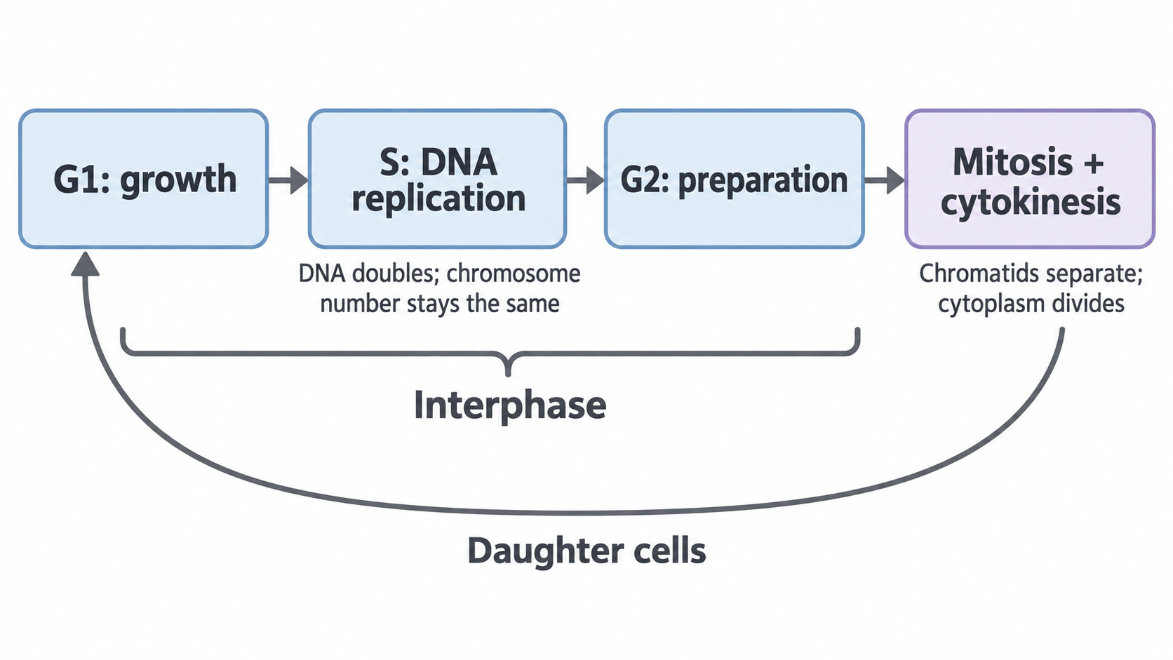
Box sizes do not represent duration.
What happens in interphase
Interphase is not a resting phase. It is a period of intense activity:
- In G1, the cell grows and synthesises proteins and organelles.
- In S phase, DNA is replicated, so each chromosome is copied.
- In G2, the cell continues to grow and synthesises the proteins needed for division.
Interphase
Interphase is the stage of the cell cycle in which DNA is replicated and the cell prepares for division.
Under a microscope you see an intact nucleus but no separate chromosomes, because the DNA is dispersed as less-condensed chromatin. DNA is present throughout, so chromosomes becoming visible later does not mean they have just been made.
Counting chromosomes and DNA
By the end of interphase, each chromosome consists of two identical sister chromatids. Each chromatid contains one DNA molecule with the same sequence, apart from copying errors, and the two remain joined at the centromere.
Count chromosomes by their centromeres, not by the arms drawn: one X shape is one replicated chromosome, not two homologous chromosomes. Homologous chromosomes are the corresponding maternal and paternal chromosomes and may carry different alleles (versions of a gene). Mitosis separates sister chromatids, not homologous pairs.
For a cell with four chromosomes before replication:
| Stage | Chromosomes in the whole cell | DNA molecules in the whole cell |
|---|---|---|
| G1 | 4 | 4 |
| End of S, G2, prophase or metaphase | 4 | 8 |
| Anaphase, before cytokinesis | 8 (4 moving to each pole) | 8 |
| Each daughter cell after cytokinesis | 4 | 4 |
Replication doubles DNA quantity without changing chromosome number, because the copies stay joined at one centromere. In anaphase the separated chromatids become chromosomes, so the count in the still-undivided cell temporarily doubles. Each new nucleus then receives the original chromosome number.
Relative DNA mass follows the same logic. If G1 DNA mass is 6 arbitrary units per cell, it rises to 12 through S, remains 12 through G2 and early mitosis, then returns to 6 per daughter cell after division.
Mitosis and cytokinesis
Every stage of mitosis looks the way it does for a reason, and those reasons are how you identify it.
Mitosis
Mitosis is nuclear division that produces two daughter nuclei containing genetic information identical to that of the parent nucleus.
Cytokinesis then usually follows, so a eukaryotic cell produces two daughter cells, each with identical copies of the DNA made during replication. The key event is the separation of the two chromatids of each chromosome.
Chromatid
A chromatid is one of two identical copies of a chromosome formed by DNA replication. The two chromatids are joined at the centromere until anaphase.
The four stages of mitosis
Mitosis is a continuous process, but it is usually described in four stages. Follow the blue chromosome across this simplified animal cell, which starts with two replicated chromosomes.
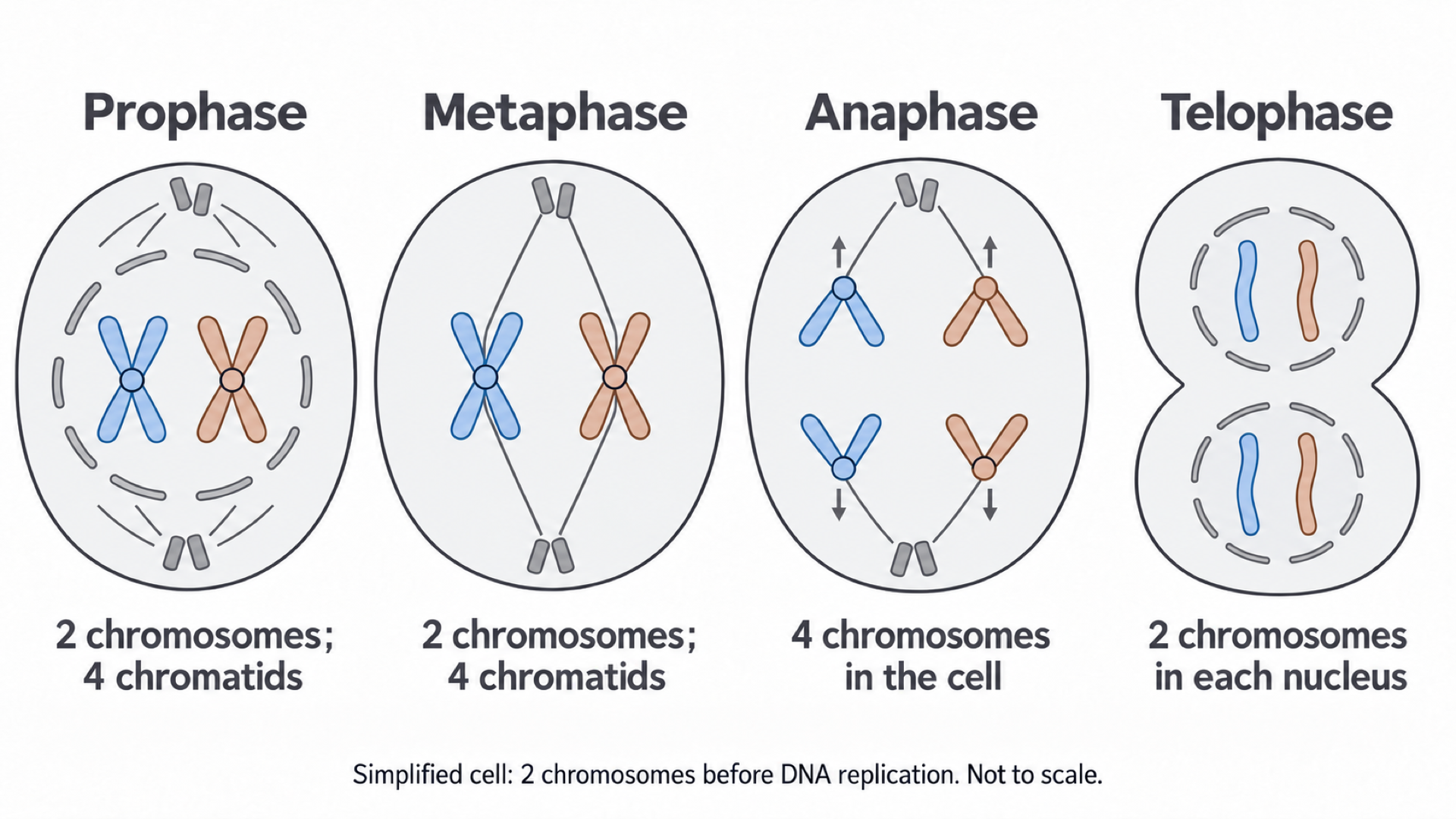
- Prophase. Chromosomes condense: coiling packs the DNA into short, thick structures that take up stain strongly, so they become visible under the light microscope as two sister chromatids joined at the centromere. Spindle fibres, protein strands that stretch between the poles, begin to form. In animal cells, centrioles (small protein cylinders that organise the spindle) move to opposite poles; root tip cells form a spindle without centrioles. The nuclear envelope breaks down, so spindle fibres can attach to centromeres in late prophase.
- Metaphase. Chromosomes line up at the equator of the cell. Spindle fibres from opposite poles are attached at the centromeres of the sister chromatids, so one copy of each chromosome can be moved to each pole.
- Anaphase. The centromeres divide, which frees the sister chromatids from each other. Spindle fibres shorten and pull them to opposite poles, a movement that requires energy from ATP. The centromere leads and the arms trail behind, which gives the V shape you see under the microscope. Once separated, each chromatid is described as a chromosome.
- Telophase. Chromosomes reach the poles, uncoil and become less visible. A nuclear envelope forms around each group, giving two genetically identical nuclei.
At anaphase the still-undivided cell holds four chromosomes, two moving to each pole, so each daughter nucleus receives two, the same as the parent.
Exam tip
To describe anaphase, state that the centromeres divide, then that chromatids move to opposite poles. Examiners report that many students miss the centromere, and writing “homologous chromosomes” here can lose the mark, because those separate in meiosis (3.4.3).
Cytokinesis
Cytokinesis divides the cytoplasm and usually overlaps late mitosis or follows it, producing two separate daughter cells.
- In animal cells, the cell-surface membrane pinches inward.
- In plant cells, vesicles form a cell plate that develops into a new cell wall, so a cell plate beginning to form marks cytokinesis.
Recognising stages in a root tip squash
AQA questions often show stained cells from a plant root tip and ask for the stage. Focus on the arrangement of chromosomes and whether the nucleus is intact.
| Stage | What you see | Why it looks like this |
|---|---|---|
| Interphase | An intact nucleus, with no separate chromosomes | The DNA is spread out as chromatin |
| Prophase | Dark, condensed threads or clumps, not lined up; nuclear envelope breaking down | Coiled chromosomes take up stain strongly; they have not yet reached the equator |
| Metaphase | A clear line of chromosomes across the equator | Spindle fibres from both poles hold the centromeres at the equator |
| Anaphase | Two groups being pulled apart, often V-shaped | Shortening fibres pull the centromeres first, so the arms trail |
| Telophase | Two groups at opposite poles; two new nuclei may be visible | Chromosomes have arrived and start to uncoil as nuclear envelopes form |
| Cytokinesis | A developing cell plate between the two new nuclei | Vesicles are building a new wall across the middle of the cell |
Interphase is usually the most common stage in a root tip squash, because cells spend much longer in interphase than in mitosis.
Growth, repair and cell numbers
Mitosis produces genetically identical cells, and three of its uses depend on that.
- Growth: a zygote (fertilised egg) divides repeatedly to produce the many cells of a multicellular organism.
- Repair and replacement: damaged or worn-out cells can be replaced with cells that have the same genes.
- Asexual reproduction: some organisms produce offspring by mitosis, so the offspring are genetically identical to the parent apart from any mutation.
Retaining a complete copy of the genome allows replacement cells to express the genes needed for the tissue's function.
Predicting cell numbers
If every cell divides once in each cycle and none dies, the number of cells doubles each cycle.
Cell number after repeated division
is the starting number of cells and is the number of consecutive cycles.
Standard form writes a number as , with .
Worked example: cells after five cycles
25 cells complete five cycles. Give the final number in standard form.
Plan: apply the formula, then convert.
Answer: cells.
Check: doubling by hand gives 50, 100, 200, 400, 800.
A correct number left out of standard form can lose a mark when a question asks for it. Reverse the calculation by dividing by to estimate a starting population.
This ideal model rests on the assumption that every cell completes every cycle; real tissues include non-dividing cells, cell death and different cycle lengths. It therefore gives a prediction, not a guaranteed tumour growth rate.
Uncontrolled division and cancer
Tumours form when the controls on mitosis fail, and many cancer drugs slow division.
Mitosis is a controlled process: genes and cell-signalling molecules regulate when a cell divides. If control is lost, cells may divide too often and form a tumour.
Tumour
A tumour is a mass of abnormal cells produced by uncontrolled cell division.
Tumours may be:
- Benign: contained in one place and not invasive.
- Malignant: invasive and able to spread to other parts of the body. Malignant tumours are cancers.
How cancer drugs slow division
Many cancer treatments are directed at controlling the rate of cell division. Drug treatment of this kind is called chemotherapy:
- Some drugs prevent DNA replication, so the cell cannot make the second copy of its DNA that mitosis needs.
- Some drugs prevent spindle fibres forming, so chromosomes cannot separate properly.
Either way, the rate of cell division falls, so the tumour grows more slowly. Cells that divide often are hit hardest, because they are more likely to be replicating DNA or forming a spindle while the drug is present.
These treatments can also affect normal tissues in which cells divide frequently, such as the gut lining, bone marrow, and hair follicles.
Binary fission
Prokaryotes have no nucleus to divide, yet each daughter cell still receives a full copy of the circular DNA.
Binary fission
Binary fission is the way prokaryotic cells divide to produce two daughter cells.
Prokaryotes do not divide by mitosis because they do not have a nucleus or linear chromosomes. Instead:
- The circular DNA replicates.
- Plasmids replicate independently of the circular DNA.
- The cytoplasm divides to produce two daughter cells. Each receives one copy of the circular DNA and a variable number of plasmids, because a cell can hold many plasmid copies and they need not be shared out equally.
Binary fission involves no chromosome condensation, spindle fibres or nuclear envelope.
| Mitosis | Binary fission | |
|---|---|---|
| Occurs in | Eukaryotic cells | Prokaryotic cells |
| Genetic material | Linear chromosomes in a nucleus | Circular DNA and plasmids |
| Spindle fibres | Yes | No |
| Named stages | Prophase to telophase | None |
| What each product receives | One copy of every chromosome | One copy of the circular DNA, plus a variable number of plasmids |
Cover the table, then list the differences from memory.
How new virus particles are made
Viruses do not divide: an infected host cell builds the new particles.
Virus
A virus is an acellular particle made of nucleic acid surrounded by a protein coat. It can replicate only inside a host cell.
Viruses are non-living and do not undergo cell division. A virus has no ribosomes or complete metabolic machinery, so after infection the host cell replicates the particles:
- Attachment proteins bind the virus to a complementary receptor on a host cell.
- The viral nucleic acid is injected into the host cell. Some viruses use another route: HIV's envelope fuses with the cell membrane (3.2.4b).
- Viral nucleic acid directs the host cell to make viral nucleic acid and proteins, using host ribosomes, energy and raw materials. Depending on the virus, host and/or viral enzymes copy its genome.
- New virus particles are assembled. Copying the genome alone is insufficient: capsid and attachment proteins must also be made and assembled with that genome.
- The new viruses leave the host cell, often when the cell lyses (bursts).
So a suitable host cell is necessary, and viral replication differs from the doubling of a bacterium.