3.1.2 - Carbohydrates
A potato cell stores starch inside a cellulose wall. Both are chains of glucose, yet one is a compact store and the other a strong wall, and the difference starts at a single carbon atom. You also learn to test for, separate and measure carbohydrates.
Monosaccharides
Every carbohydrate in this lesson is built from small sugar units called monosaccharides.
Monosaccharide
A monosaccharide is a single sugar unit and the monomer from which larger carbohydrates are made.
You need three: glucose, galactose and fructose. They are small and soluble, and all three are , fitting the general formula . Glucose is a major respiratory substrate and the monomer of starch, glycogen and cellulose.
The two isomers of glucose
Glucose has two isomers, alpha-glucose (α-glucose) and beta-glucose (β-glucose): they share the molecular formula but differ in the arrangement of their atoms. In the diagram, compare the highlighted OH groups on carbon 1, the corner beside the ring oxygen.
[DIAGRAM: asset_name: 1.2 - Carbohydrates - Diagram 1; asset_slug: 1.2 - Carbohydrates - Diagram 1; recommended_method: retained_png; caption: alpha-glucose and beta-glucose ring structures with the OH group on carbon 1 labelled.; description: alpha-glucose and beta-glucose ring structures with the OH group on carbon 1 labelled.]
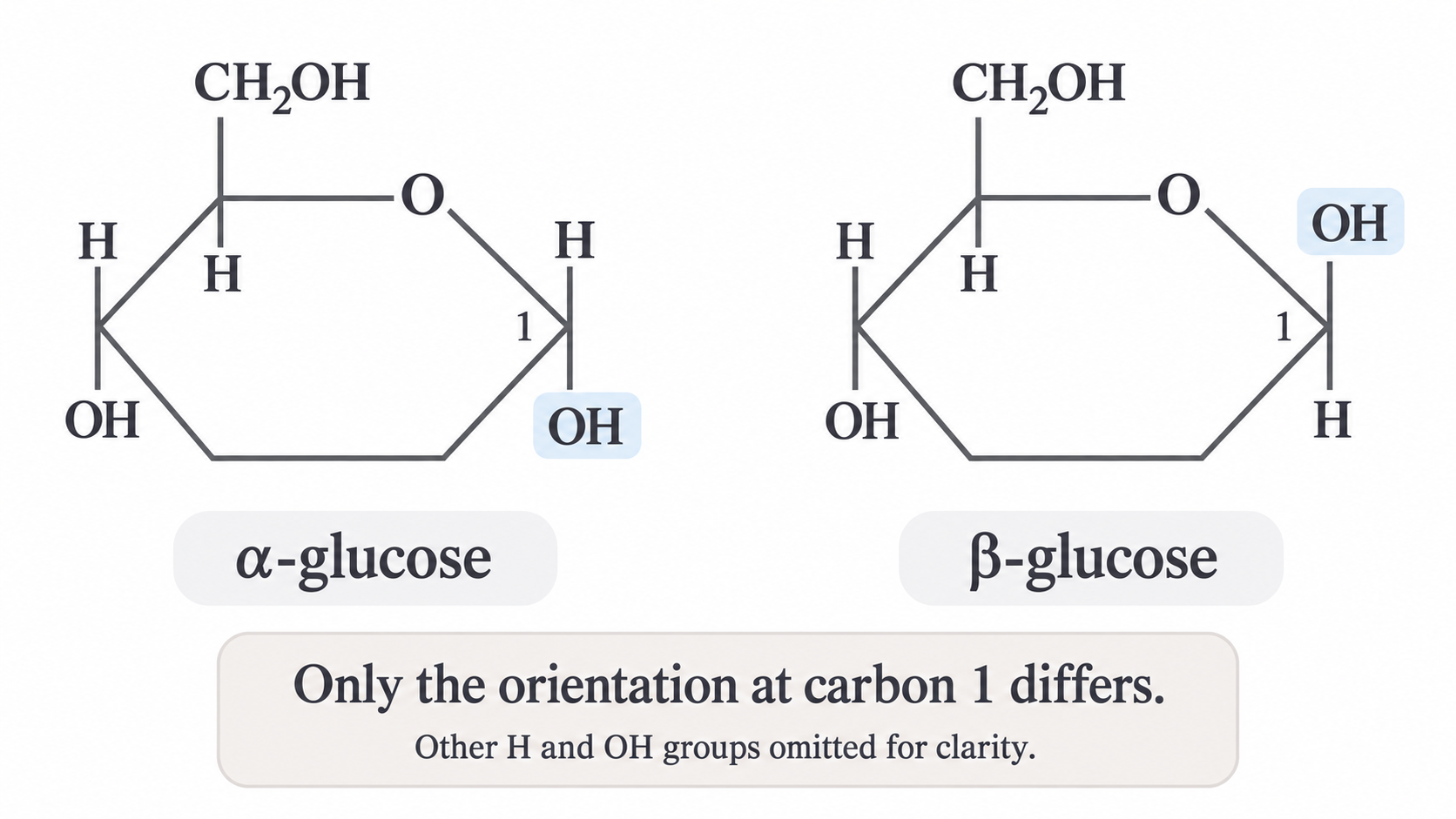
In this conventional drawing, only carbon 1 differs:
- In alpha-glucose, the hydroxyl (OH) group on carbon 1 is below the ring.
- In beta-glucose, the hydroxyl group on carbon 1 is above the ring.
Rotating a drawing does not change the isomer, so for a rotated ring compare the carbon 1 OH with the on carbon 5: opposite sides means alpha, the same side means beta. That one swap decides whether glucose chains coil or run straight.
Disaccharides and glycosidic bonds
Two glucose molecules join to make maltose, losing a molecule of water.
This is the condensation reaction from 3.1.1, and it joins any two monosaccharides:
- The OH from one sugar and the H from an OH group on the other form one water molecule.
- The oxygen atom left behind links the two sugars in a glycosidic bond.
Glycosidic bond
A glycosidic bond is the covalent bond formed between two monosaccharides in a condensation reaction.
In the maltose diagram, the oxygen bridge joins carbon 1 of one glucose to carbon 4 of the other. In the label α-1,4, the numbers name those carbons and α gives the bond's orientation at carbon 1.
[DIAGRAM: asset_name: 1.2 - Carbohydrates - Diagram 2; asset_slug: 1.2 - Carbohydrates - Diagram 2; recommended_method: retained_png; caption: Simplified maltose structure with an alpha-1,4 glycosidic linkage and the net condensation word equation.; description: Simplified maltose structure with an alpha-1,4 glycosidic linkage and the net condensation word equation.]
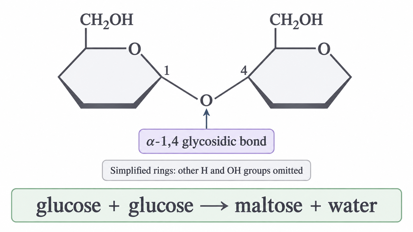
A disaccharide is made from two monosaccharides. You need these three:
| Disaccharide | Monosaccharides joined |
|---|---|
| Maltose | glucose + glucose |
| Sucrose | glucose + fructose |
| Lactose | glucose + galactose |
Hydrolysis is the reverse: water is used to break the glycosidic bond, releasing the monosaccharides. Those released in the small intestine are small enough to be absorbed through its wall.
Storage polysaccharides
A cell packed with dissolved glucose would take in water by osmosis.
Starch and glycogen hold the same glucose in a form that does not.
Polysaccharides form when many glucose units join by condensation reactions. Starch and glycogen are both made from alpha-glucose, but their chains are arranged differently. In the diagram, each circle is one glucose unit.
[DIAGRAM: asset_name: 1.2 - Carbohydrates - Storage polysaccharides; asset_slug: 1.2 - Carbohydrates - Storage polysaccharides; recommended_method: imagegen; caption: Coiled unbranched amylose, branched amylopectin and more highly branched glycogen.; description: Coiled unbranched amylose, branched amylopectin and more highly branched glycogen.]
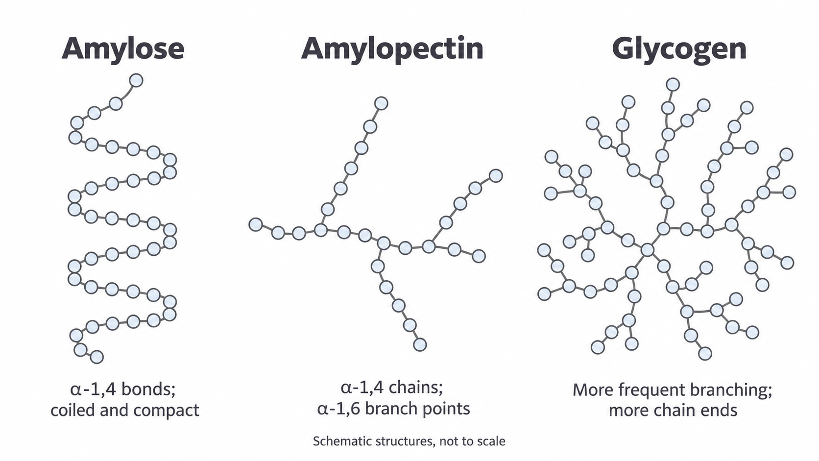
- Amylose, in plant starch, is an unbranched, coiled chain with 1,4 glycosidic bonds.
- Amylopectin, also in starch, is branched, with 1,4 glycosidic bonds in the chains and 1,6 glycosidic bonds at branch points.
- Glycogen, the store in animals, is similar to amylopectin but more highly branched.
Why these structures store glucose well
Starch and glycogen are:
- Insoluble, so they add no dissolved solute particles to lower water potential. Water moves by osmosis towards lower water potential, so they draw no water into the cell.
- Large, so they do not diffuse out of cells.
- Compact, because of coiling and branching, so a lot can be stored in a small space.
- Branched, giving many chain ends where enzymes can act at once, so hydrolysis releases glucose rapidly.
Glycogen's extra branching gives it more ends than starch, so it releases glucose faster when respiration demand rises, as in an active muscle cell. Starch is a good long-term store in plant cells.
In an answer, follow each feature with its consequence: "insoluble, so it does not lower water potential", not just "insoluble".
Cellulose
Cellulose is also a polysaccharide of glucose, yet it forms strong fibres in plant cell walls.
The difference begins with its monomer: beta-glucose, not alpha-glucose.
Why beta-glucose gives straight chains
Each glycosidic bond in these chains joins the OH on carbon 1 of one unit to the OH on carbon 4 of the next. Carbon 4 is the left-hand corner of the ring in the glucose diagram, and its OH is below the ring in both isomers.
- In alpha-glucose, the carbon 1 OH is also below the ring, so neighbouring units join the same way up and the chain coils.
- In beta-glucose, the carbon 1 OH is above the ring, so the two groups can only meet in a condensation reaction if one unit flips.
So each beta-glucose unit is rotated 180 degrees relative to the next, giving long, straight, unbranched chains.
From chains to a strong wall
In the diagram, each arrow is a glucose unit. Solid lines are covalent bonds within a chain; dotted lines are hydrogen bonds between chains.
[DIAGRAM: asset_name: 1.2 - Carbohydrates - Diagram 3; asset_slug: 1.2 - Carbohydrates - Diagram 3; recommended_method: retained_png; caption: cellulose chains with alternate beta-glucose orientation, hydrogen bonds between chains, and grouping into microfibrils.; description: cellulose chains with alternate beta-glucose orientation, hydrogen bonds between chains, and grouping into microfibrils.]
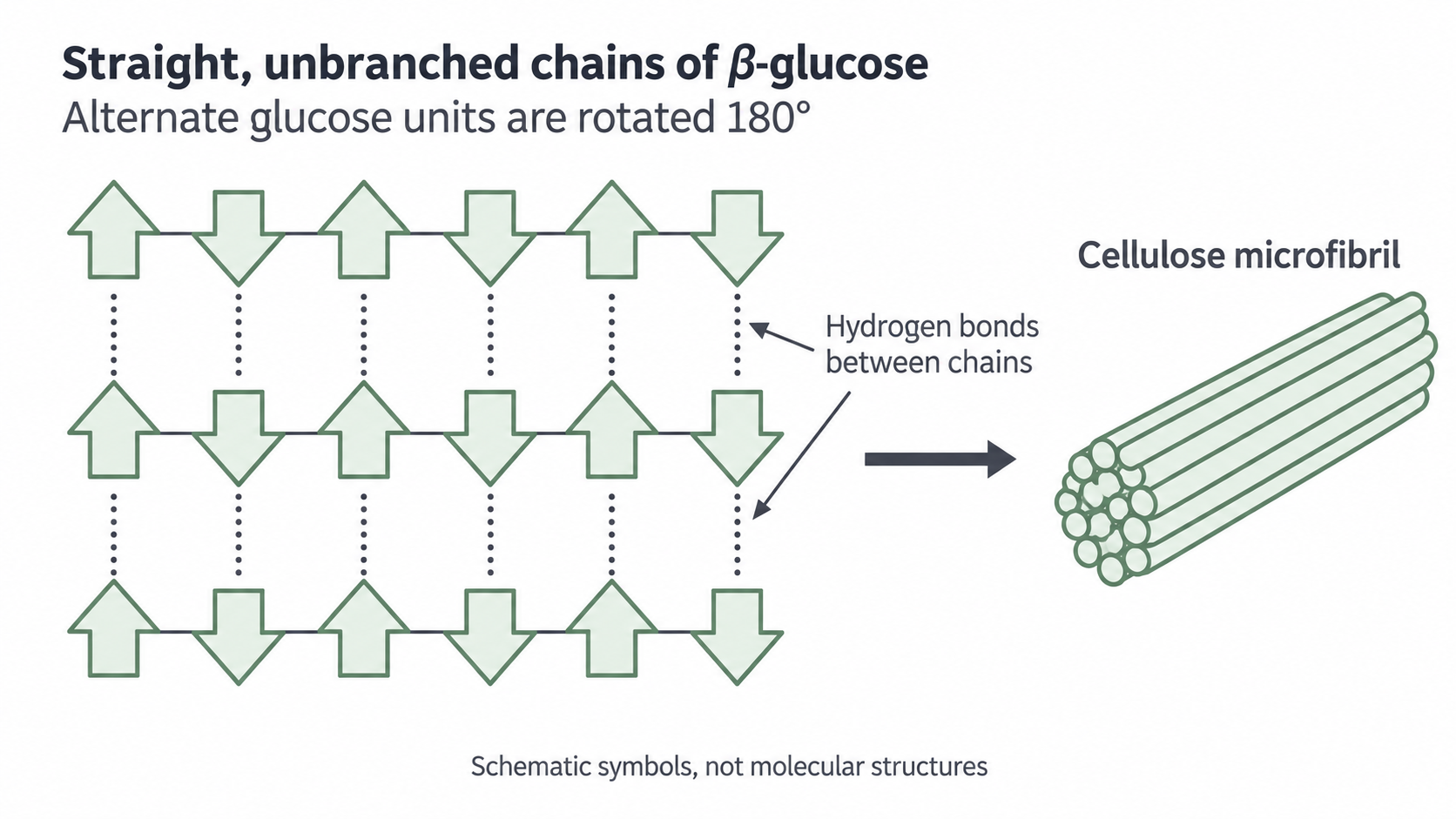
- Straight chains pack closely side by side, so many hydrogen bonds form between them.
- Chains group into microfibrils and microfibrils into fibres, giving the cell wall high tensile strength (resistance to being pulled apart).
- When water enters the cell by osmosis, its contents press against the wall, and the wall resists bursting.
- The cell becomes turgid (firm), and turgid cells help support non-woody parts of the plant.
Watch out
A single hydrogen bond is weaker than a covalent glycosidic bond. Cellulose is strong because many hydrogen bonds act together, resisting the chains sliding apart.
The three polysaccharides compare like this:
| Starch | Glycogen | Cellulose | |
|---|---|---|---|
| Monomer | Alpha-glucose | Alpha-glucose | Beta-glucose |
| Glycosidic bonds | 1,4 and 1,6 | 1,4 and more 1,6 | 1,4 only |
| Shape | Coiled amylose, branched amylopectin | Highly branched | Straight, unbranched, parallel chains |
| Found in | Plant cells | Animal cells | Plant cell walls |
| Function | Carbohydrate store | Carbohydrate store | Strength and support |
Cover the table, then explain the differences from memory.
Biochemical tests for carbohydrates
Simple colour tests show which carbohydrates a food contains.
Crush a solid sample with distilled water, filter if needed, and use a separate portion for each test so reagents do not interfere. A negative result means the substance was not detected under the test conditions, not that none is present.
Reducing and non-reducing sugars
A reducing sugar reduces the blue copper(II) ions in Benedict's reagent, forming a precipitate of red copper(I) oxide. Glucose, fructose, galactose, maltose and lactose are all reducing sugars. To test for one:
- Add Benedict's reagent to the sample.
- Heat in a hot water bath for a standard time, such as five minutes, using eye protection and a test-tube holder.
- Observe the colour. Blue is negative. A green, yellow, orange or brick-red precipitate is positive.
More reducing sugar forms more precipitate and leaves less blue solution, so the colour moves from green towards brick-red. Even with volumes, temperature and heating time kept the same, this is only a rough guide to concentration, and Benedict's detects reducing sugars other than glucose.
Exam tip
Write "heat with Benedict's reagent". AQA's 2024 mark scheme ignored "warm" and an unqualified "water bath", and rejected heating with acid.
Sucrose, unlike maltose and lactose, is a non-reducing disaccharide. Benedict's reagent stays blue until hydrolysis releases its reducing monosaccharides:
- Test an untreated portion with Benedict's reagent. If it stays blue, continue with a fresh portion.
- Heat the fresh portion with dilute hydrochloric acid to hydrolyse glycosidic bonds.
- Cool it, then carefully neutralise the acid with sodium hydrogencarbonate. Neutralisation removes the acid, which would otherwise stop Benedict's reagent working; it does not split the sugar.
- Add Benedict's reagent and heat again.
A positive result after an initially negative test supports a non-reducing sugar such as sucrose, though other carbohydrates in a food can hydrolyse too. If the first test was already positive, a positive result afterwards cannot show whether non-reducing sugar is also present without a quantitative comparison.
Starch and controls
Add iodine in potassium iodide solution to a separate sample at room temperature, without heating. Yellow-brown turning blue-black indicates starch; a negative result stays yellow-brown.
In any of these tests, a known positive control checks the reagent and method, and distilled water as a negative control reveals contaminated reagents or equipment.
Identifying monosaccharides
Glucose, galactose and fructose are all reducing sugars, so Benedict's reagent cannot tell them apart.
Chromatography can: it separates a mixture, and known standards identify the components.
Running the chromatogram
- Draw a pencil baseline near the bottom of the chromatography paper, because ink would dissolve in the solvent and run.
- Place small spots of the unknown and of known glucose, galactose and fructose solutions on it. Let each dry; repeated small applications concentrate a dilute sample without blurring the spot.
- Stand the paper in a suitable solvent with the baseline above the surface, otherwise the sugars would dissolve straight into the solvent.
- Cover the container, which stops solvent evaporating from the paper, and let the solvent rise.
- Remove the paper before the solvent reaches the top and immediately mark the solvent front in pencil, because it fades as the solvent evaporates.
- Apply a suitable locating reagent to reveal the colourless sugars, following its safety instructions.
Different sugars move different distances because their affinities for the mobile solvent and the stationary phase (the paper) differ. A sugar more attracted to the solvent than to the paper travels further.
Identifying the spots
Compare the unknown's spots with standards developed on the same paper, then calculate:
Rf value
Both distances use the same units, so has no unit.
If the solvent front moves 80 mm and a glucose standard moves 32 mm, its is . An unknown spot at the same position is consistent with glucose under those conditions, but it is not unique proof, because different compounds can travel together. A second solvent or method strengthens identification.
values depend on the solvent, so compare them only within one solvent.
Measuring glucose concentration
A Benedict's colour gives only a rough concentration.
A colorimeter gives a number, and a calibration curve relates that signal to known concentrations, so an unknown can be estimated by comparison.
Making a dilution series
A dilution reduces concentration while preserving the amount of solute taken from the stock:
Dilution
Subscript 1 is the stock and subscript 2 is the diluted solution. Use consistent units.
To make 10 cm³ of 2 mg cm⁻³ glucose from a 10 mg cm⁻³ stock, take cm³ of stock and add 8 cm³ of water. The same calculation gives a full series, including a zero-glucose blank:
| Glucose (mg cm⁻³) | Stock (cm³) | Water (cm³) |
|---|---|---|
| 0 | 0 | 10 |
| 2 | 2 | 8 |
| 4 | 4 | 6 |
| 6 | 6 | 4 |
| 8 | 8 | 2 |
| 10 | 10 | 0 |
Mix each thoroughly, using volumetric apparatus suited to the required precision.
Building and using the curve
One possible method measures the blue solution left after the Benedict's reaction:
- Add excess Benedict's reagent to each standard, so glucose limits the reaction. Use fixed volumes and identical heating times and temperatures.
- After cooling, remove the precipitate by the same filtration or centrifugation procedure, because suspended solid would scatter light.
- Use a suitable filter, usually red, because a blue solution absorbs red light strongly, and zero the colorimeter with the optical blank, usually distilled water in a cuvette.
- Measure each absorbance with repeats, keeping cuvettes clean, consistently oriented and filled to the same level.
- Plot glucose concentration on the horizontal axis and mean absorbance on the vertical axis, then draw the curve, which need not be straight. AQA rejects absorbance on the x-axis.
- Treat the unknown identically, so that concentration is the only difference, and read its value from the curve.
Watch out
Here absorbance falls as concentration rises, because more reducing sugar consumes more blue copper(II) ions. Your zero-glucose blank goes through the whole method and gives the highest absorbance. It is not the optical blank.
Interpolate within the standards' range, because the curve is only known there. If the unknown falls outside it, dilute and repeat instead of extrapolating, then multiply the value from the curve by the dilution factor.
Worked example: correcting for dilution
Question. A sample is diluted one part sample to four parts water. The diluted sample reads 0.80 mg cm⁻³. Find the original concentration.
Plan. Find the dilution factor from the total parts, then multiply the reading by it.
Answer. The original contained 4.0 mg cm⁻³.
Check. The original must be more concentrated than its diluted sample, and 4.0 is greater than 0.80.
This method estimates glucose only if other reducing substances do not interfere; a biological mixture may require a glucose-specific assay.