6.33P-6.35P - Medical radiation and PET
Compare two ways to target a tumour, then follow the separate diagnostic process from a radioactive tracer to a PET image. The source’s position, emitted radiation and half-life determine how it can be used.
Internal and external radiotherapy
Radiotherapy uses ionising radiation to damage the DNA of tumour cells so that they die or can no longer divide. The central problem is to give the tumour a large enough dose while keeping the dose to surrounding healthy tissue as low as practicable.
In external-beam radiotherapy, a source outside the body directs penetrating radiation towards the tumour. Modern treatment may use machine-produced high-energy X-rays or particles; it does not always use a radioactive isotope. Beams can enter from several directions and overlap at the tumour, so the target receives a larger total dose than any one region of healthy tissue along a single beam path. A machine-produced beam can be switched off; a radioactive-source beam is stopped by closing shielding or moving the source. In either arrangement, no radioactive material remains in the patient after treatment.
In internal radiotherapy, a radiation source is placed inside the body, in or very close to the tumour. A common example is brachytherapy, in which a small sealed source is inserted temporarily or implanted. Because the source is close to the target, a high local dose can be delivered while the dose falls away through nearby tissue. Some internal treatments instead use a radioactive substance that is taken up preferentially by a target organ.
[DIAGRAM: asset_name: 28_1PH0-P1-06E_6.28P-6.35P - Radiation uses, hazards and medicine - diagram 02; asset_slug: edexcel-gcse-physics-6-28p-6-35p-radiation-uses-hazards-medicine_diagram_02; recommended_method: image_gen; description: paired monochrome medical diagram comparing external beams converging on a tumour with a sealed brachytherapy source positioned inside or beside the tumour]
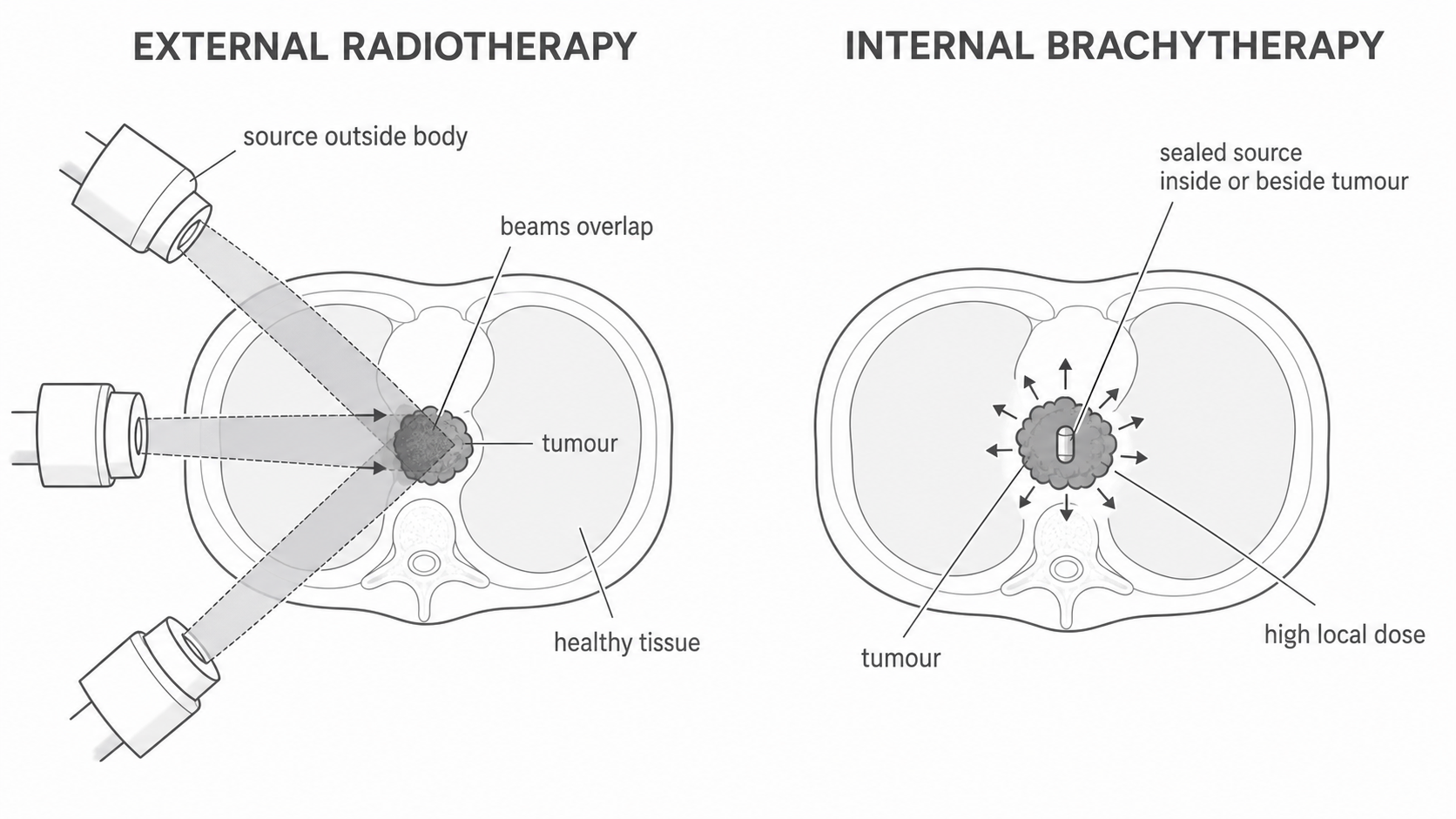
| Feature | External treatment | Internal treatment |
|---|---|---|
| Source position | outside the patient | in or close to the tumour |
| Dose pattern | beams pass through some healthy tissue before and after the target | high dose is concentrated close to the internal source |
| Control | beam direction, shape, intensity and exposure time are controlled; beam can be stopped | source position, activity and time in the body are controlled; a temporary source can be removed |
| Useful situation | can reach tumours at many positions without inserting a source | can spare more distant healthy tissue when the tumour is suitable and accessible |
| Limitation | healthy tissue along beam paths receives some dose | placement may be invasive and is not suitable for every tumour; the source must be securely contained and accounted for |
Both methods can damage healthy as well as cancerous cells. Neither is automatically better: the tumour's position, size and type determine which route can deliver the best-localised dose. A sealed brachytherapy source is deliberately contained; it should not be confused with an accidental spill of radioactive material.
PET and diagnostic tracers
Diagnostic tracers reveal function rather than deliberately destroying tissue. A small amount of radioactive substance is attached to, or behaves like, a material used by the body. A detector outside the body maps where the tracer travels or collects. For example, different uptake in an organ can reveal unusual blood flow or metabolism.
A PET scan uses a tracer containing a positron-emitting isotope. The process is a causal chain:
Continue this lesson.
You’re previewing NovaLearn without an account. Create a free account to read the rest.
Create free account