2.91-2.92 - The eye - structure, focusing and light response
The eye is a sense organ that acts as a receptor for light. It does more than let light in: it focuses light onto receptor cells in the retina, then sends electrical impulses to the central nervous system. The same eye also adjusts when objects are near or distant, and when light intensity changes.
The eye as a receptor
A stimulus is a change in the environment. For the eye, the stimulus is light. The eye acts as a receptor because light-sensitive cells in the retina detect light and start electrical impulses.
Receptor
A receptor is a cell or sense organ that detects a stimulus and starts a response.
The parts of the eye work together so light can be detected clearly:
| Structure | Function |
|---|---|
| Sclera | Tough outer coat that protects the eye and helps keep its shape |
| Cornea | Transparent front surface that lets light in and begins to refract it |
| Iris | Ring of muscle that controls the size of the pupil |
| Pupil | Hole that allows light to enter the eye |
| Lens | Transparent structure that changes shape to focus light |
| Ciliary muscles | Muscles that change tension in the suspensory ligaments |
| Suspensory ligaments | Fibres that connect the ciliary body to the lens and alter lens shape |
| Retina | Light-sensitive layer containing receptor cells |
| Optic nerve | Carries electrical impulses from the retina to the brain |
Light enters through the cornea and pupil, passes through the lens, and reaches the retina. Receptor cells in the retina convert the light stimulus into electrical impulses. These impulses travel along the optic nerve to the brain, where the information is processed.
[DIAGRAM: asset_name: Eye structure labelled schematic; asset_slug: b37_the_eye_structure_focusing_and_light_response__diagram_01; recommended_method: deterministic_drawing; description: A monochrome labelled cross-section of the eye showing sclera, cornea, iris, pupil, lens, ciliary muscles, suspensory ligaments, retina and optic nerve, with arrows showing light entering and impulses leaving.]
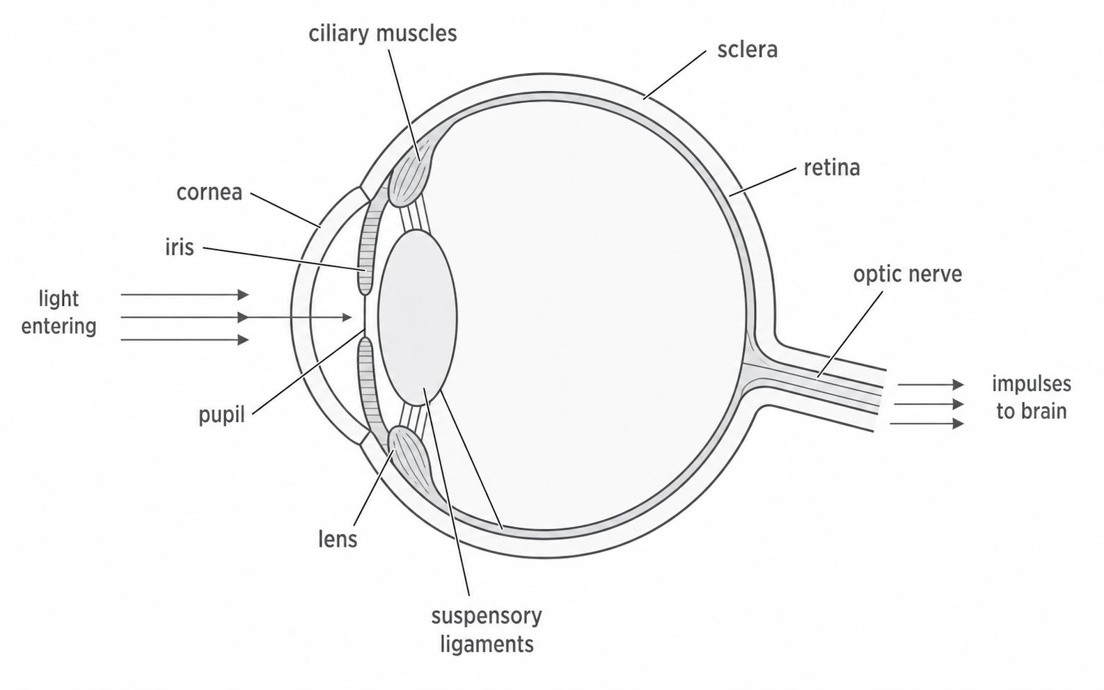
Focusing light on the retina
For vision to be clear, light from an object must be focused onto the retina. If the light is focused in front of or behind the retina, the image reaching the receptor cells is blurred.
The cornea and lens both refract light. In this lesson, the important adjustable part is the lens. The lens can change shape, which changes how strongly it refracts light.
Accommodation
Accommodation is the change in lens shape that allows the eye to focus on objects at different distances.
A thin lens refracts light less strongly. A thicker, more rounded lens refracts light more strongly. This matters because light from near objects needs more refraction to be focused on the retina than light from distant objects.
The retina stays in the same place at the back of the eye. The eye solves the focusing problem by changing the shape of the lens, not by moving the retina.
Near and distant objects
Accommodation depends on the ciliary muscles, suspensory ligaments and lens. These three parts form a simple control chain:
ciliary muscles -> suspensory ligaments -> lens shape -> focus on retina
When looking at a distant object, the ciliary muscles relax. This makes the suspensory ligaments tight. The tight ligaments pull on the lens, making it thinner and less rounded. A thinner lens refracts light less strongly, which is enough for distant light rays to focus on the retina.
When looking at a near object, the ciliary muscles contract. This reduces the tension in the suspensory ligaments. The lens becomes thicker and more rounded. A thicker lens refracts light more strongly, so light from the near object can still be focused on the retina.
Continue this lesson.
You’re previewing NovaLearn without an account. Create a free account to read the rest.
Create free account