2.67-2.69 - Blood vessels, circulation and coronary heart disease
Blood vessels are not just tubes: their walls, lumens and valves match the pressure and job of the blood they carry. This lesson links those vessel adaptations to the whole circulation map, including the vessels serving the lungs, liver and kidneys. It also shows how risk factors can increase the chance of coronary heart disease by affecting the coronary arteries that supply the heart muscle.
Coronary heart disease risk
The heart muscle needs its own blood supply because its cells need glucose and oxygen for aerobic respiration. The vessels that supply the heart muscle are the coronary arteries. If these arteries become narrowed or blocked, less oxygen reaches the heart muscle, so the muscle cannot respire aerobically at the rate it needs.
Coronary heart disease
Coronary heart disease is a disease in which the coronary arteries become narrowed or blocked, reducing the blood and oxygen supply to the heart muscle.
In coronary heart disease, fatty material called atheroma can build up in the wall of a coronary artery. This narrows the lumen, so less blood can flow through. If a blood clot forms and blocks the narrowed artery, the heart muscle beyond the blockage may be starved of oxygen.
[DIAGRAM: asset_name: coronary_artery_narrowing; asset_slug: b28_blood_vessels_circulation_and_coronary_heart_disease__diagram_01; recommended_method: deterministic_drawing; description: Monochrome comparison of a normal coronary artery cross-section and a narrowed coronary artery with atheroma reducing lumen size, plus an arrow showing reduced oxygen delivery to heart muscle.]
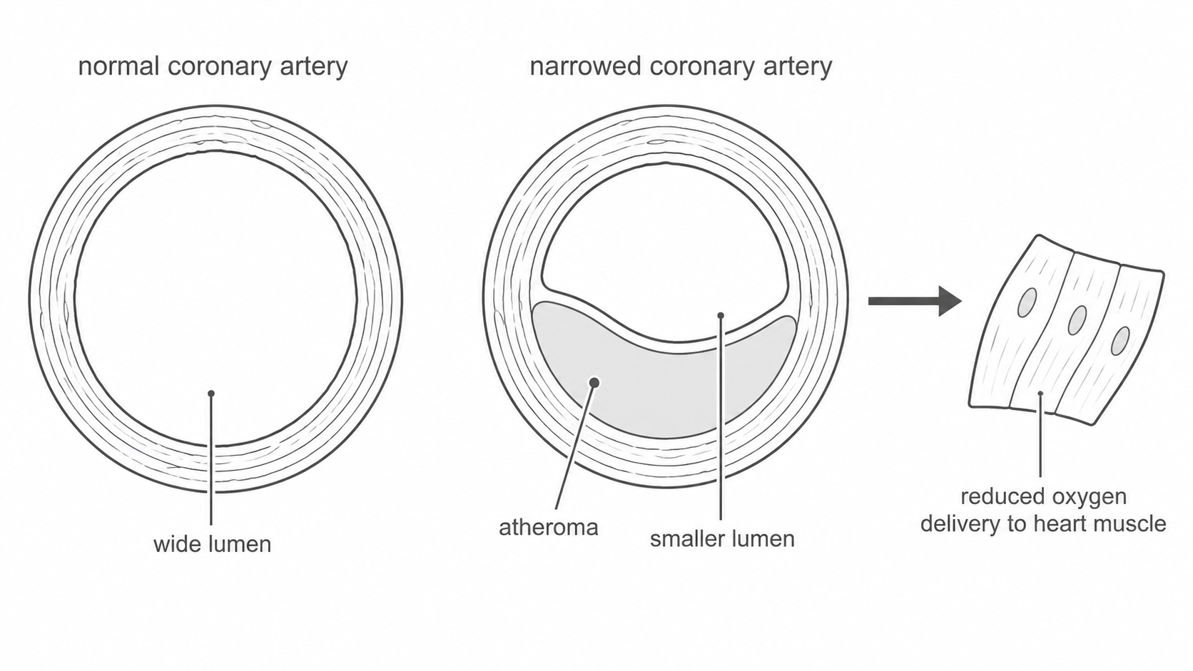
The specification asks how factors may increase the risk of developing coronary heart disease. A risk factor does not guarantee that a person will develop the disease. It makes the disease more likely, often by increasing atheroma build-up, increasing blood pressure, damaging artery linings, increasing clot risk, or increasing the work the heart must do.
Important risk factors include:
- a diet high in saturated fat or cholesterol, which can increase fatty deposits in artery walls
- high blood pressure, which puts more strain on the heart and can damage artery linings
- smoking, because chemicals in smoke can damage coronary arteries and increase the risk of blood clots
- lack of exercise, which is linked with higher blood pressure, obesity and a less efficient cardiovascular system
- obesity, which is often linked with high blood pressure, diabetes and high blood cholesterol
- diabetes, because high blood glucose can damage blood vessels
- stress, because it can raise blood pressure and may also be linked to smoking, overeating or poor sleep
- inherited factors or family history, because some people inherit a higher risk
Good exam answers do not just list factors. They link each factor to a biological effect. For example, "smoking increases CHD risk" is weaker than "smoking can damage the lining of coronary arteries and increase the chance of blood clots, so the artery is more likely to become blocked."
Arteries: high-pressure vessels
Arteries carry blood away from the heart. This is the definition to use, even when the oxygen content is unusual. Most systemic arteries carry oxygenated blood, but the pulmonary artery carries deoxygenated blood from the heart to the lungs.
Arteries receive blood at high pressure when ventricles contract. Their structure matches this function:
- thick muscular walls help withstand and control high pressure
- elastic tissue lets the artery wall stretch and recoil after each heartbeat, helping keep blood moving
- a relatively narrow lumen helps maintain high pressure
- no valves are needed along most arteries because the pressure is high enough to keep blood moving away from the heart
The elastic recoil matters. If an artery wall could only stretch and not recoil, the blood would surge and then slow too much between heartbeats. Recoil helps smooth the flow so tissues continue to receive blood between contractions.
Common exam trap: "arteries carry oxygenated blood" is not a reliable definition. Use "arteries carry blood away from the heart." Then add oxygen content only for the named vessel. For example, the aorta carries oxygenated blood away from the heart to the body, while the pulmonary artery carries deoxygenated blood away from the heart to the lungs.
Veins and capillaries
Veins carry blood towards the heart. Blood in veins is at much lower pressure than blood in arteries because it has already passed through arterioles and capillary networks. Their structure matches this lower-pressure return journey:
- thinner walls than arteries because the pressure is lower
- less muscle and elastic tissue than arteries
- a wide lumen, which reduces resistance to flow
- valves, especially in limb veins, to prevent backflow and keep blood moving towards the heart
Capillaries are the exchange vessels. They link the smallest arteries to the smallest veins through networks in tissues. Their job is not to carry blood quickly over long distances, but to allow substances to move between the blood and cells.
Capillaries are adapted for exchange because:
- their walls are one cell thick, giving a short diffusion distance
- they are very narrow, so red blood cells pass close to the capillary wall
- they form dense networks, giving a large surface area for exchange
- blood flows slowly through them, giving time for oxygen, carbon dioxide, glucose, urea and other substances to move
Continue this lesson.
You’re previewing NovaLearn without an account. Create a free account to read the rest.
Create free account