2.65-2.66 - Heart structure and heart-rate control
The heart is a muscular pump that keeps blood moving through the lungs and around the body. Its structure matters because each chamber, valve and wall helps blood move in the correct direction. Heart rate then changes when the body needs blood to be pumped faster, especially during exercise or when adrenaline is released.
The heart as a double pump
The heart is not one open bag of blood. It is a double pump with a right side and a left side working at the same time.
Double pump
A heart with two pumping sides: the right side pumps blood to the lungs, and the left side pumps blood to the body.
The heart has four chambers:
| Chamber | Main job |
|---|---|
| Right atrium | Receives deoxygenated blood from the body |
| Right ventricle | Pumps deoxygenated blood to the lungs |
| Left atrium | Receives oxygenated blood from the lungs |
| Left ventricle | Pumps oxygenated blood to the body |
The atria are the upper chambers. They receive blood and pass it down into the ventricles. The ventricles are the lower chambers. They have thicker muscular walls because they pump blood out of the heart.
The septum is the muscular wall between the left and right sides of the heart. It keeps oxygenated blood on the left side separate from deoxygenated blood on the right side. This separation means blood going to the body has a high oxygen concentration.
The left ventricle has the thickest wall. It must pump blood all the way around the body, so it contracts with higher pressure than the right ventricle, which only pumps blood to the nearby lungs.
[DIAGRAM: asset_name: Heart structure labelled schematic; asset_slug: b27_heart_structure_and_heart_rate_control__diagram_01; recommended_method: deterministic_drawing; description: A simplified labelled heart-flow schematic showing the four chambers, septum, thicker left ventricle wall, main vessels, valves and one-way blood-flow arrows through lungs and body. The patient's right side is shown on the viewer's left; separate muted colours distinguish deoxygenated and oxygenated blood.]
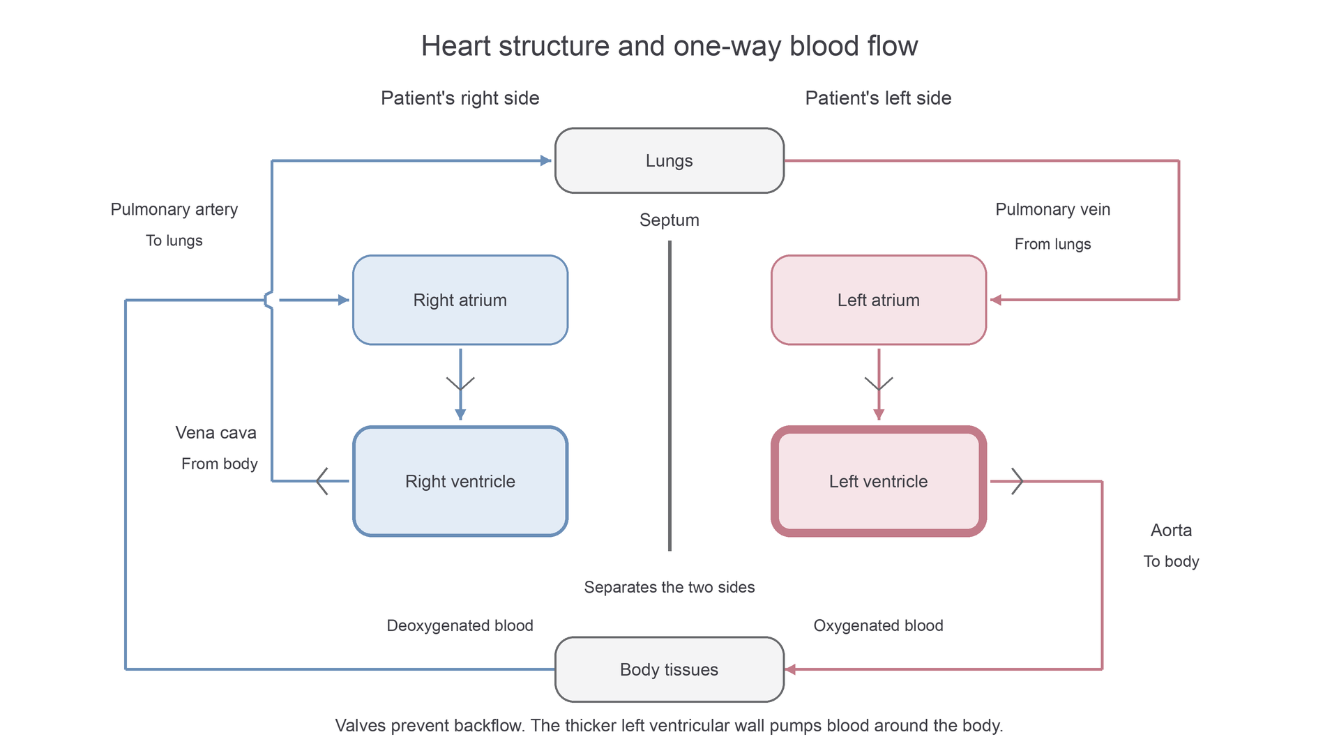
One-way flow through the heart
Blood must flow through the heart in one direction. Valves make this possible. They open when blood is moving the correct way and close to prevent backflow.
The route is:
- Deoxygenated blood from the body enters the right atrium through the vena cava.
- It passes through a valve into the right ventricle.
- The right ventricle pumps it through the pulmonary artery to the lungs.
- In the lungs, blood gains oxygen and loses carbon dioxide.
- Oxygenated blood returns through the pulmonary vein to the left atrium.
- It passes through a valve into the left ventricle.
- The left ventricle pumps it through the aorta to the body.
The pulmonary artery is an important exception: it is an artery, so it carries blood away from the heart, but it carries deoxygenated blood. The pulmonary vein is also an exception: it is a vein, so it carries blood towards the heart, but it carries oxygenated blood.
Think of the valves as direction keepers, not pumps. The heart muscle creates the pressure; the valves stop that pressure pushing blood backwards.
How a heartbeat moves blood
A heartbeat is a repeated pattern of heart muscle relaxing and contracting.
When the heart muscle relaxes, the chambers fill with blood. Blood returning from the body enters the right atrium, and blood returning from the lungs enters the left atrium.
Continue this lesson.
You’re previewing NovaLearn without an account. Create a free account to read the rest.
Create free account