2.46-2.47 - Human breathing system and ventilation
Breathing is not the same as respiration. Respiration is the chemical release of energy in cells; breathing is the physical movement of air into and out of the lungs. This lesson focuses on the structure of the human thorax and how muscles change its volume to ventilate the lungs.
The thorax is a moving cavity
The thorax is the chest region. It contains the lungs and the structures that help move air during breathing. It works like a movable cavity rather than like a pump inside the lungs, because the lungs themselves do not have muscles that actively pull air in.
Thorax
The chest cavity containing the lungs, protected by the ribs and moved by the intercostal muscles and diaphragm.
The main structures you need to recognise are:
| Structure | Position or role in the thorax |
|---|---|
| Ribs | Curved bones around the thorax that protect the lungs and help change chest volume. |
| Intercostal muscles | Muscles between the ribs. Their contractions move the rib cage during ventilation. |
| Diaphragm | A sheet of muscle below the lungs, separating the thorax from the abdomen. |
| Pleural membranes | Thin membranes around the lungs and lining the inside of the thorax; pleural fluid reduces friction as the lungs move. |
[DIAGRAM: asset_name: Human breathing system and ventilation - diagram 01; asset_slug: b20_human_breathing_system_and_ventilation__diagram_01; recommended_method: deterministic_drawing; description: Monochrome labelled front-view thorax diagram showing ribs, intercostal muscles, diaphragm, trachea, left and right bronchi, bronchioles, alveoli and pleural membranes.]
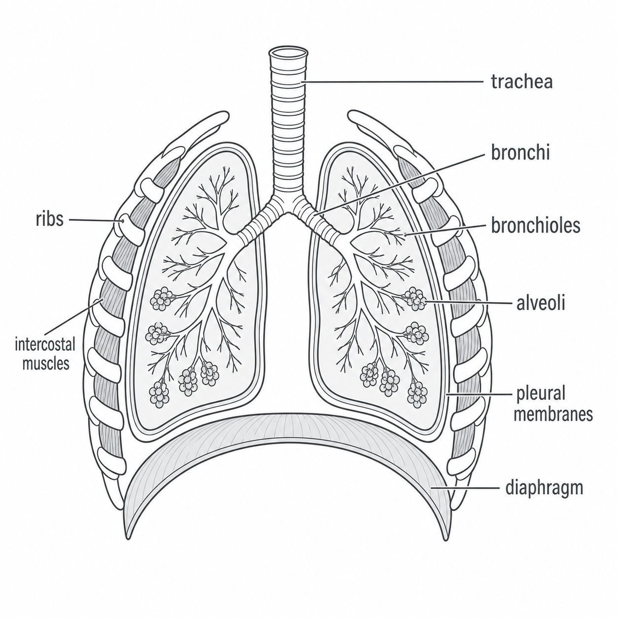
A useful mental model is: ribs and intercostal muscles change the width of the thorax; the diaphragm changes its height; pleural membranes help the lungs follow these movements smoothly.
The air pathway through the lungs
Air enters and leaves through a branching system of tubes. The order matters, because exam questions often ask students to label or complete the pathway.
The pathway is:
trachea -> bronchi -> bronchioles -> alveoli
The trachea is the windpipe. It carries air down into the chest before it divides into two bronchi, one leading into each lung. Inside the lungs, each bronchus branches repeatedly into smaller bronchioles. The bronchioles end at tiny air sacs called alveoli.
Alveolus
A tiny air sac in the lungs. The plural is alveoli.
In this lesson, you only need the alveoli as part of the thorax structure and air pathway. How alveoli are adapted for gas exchange is a separate specification point in the next cluster.
If you are tracing air movement, start with the trachea and follow the branches down to the bronchi, bronchioles and alveoli.
Continue this lesson.
You’re previewing NovaLearn without an account. Create a free account to read the rest.
Create free account