3.1.3, 3.1.7 - Brain structure and aggression
Brain functioning can help explain why a person finds it difficult to regulate a response to provocation. This lesson uses 3.1.3 to connect brain structures with aggression, and the brain-injury element of 3.1.7 to examine why patients can have different outcomes. The aim is to build an evidence-based explanation without treating a scan or an injury as a prediction of inevitable violence.
Brain structures and their interacting functions
Different brain structures contribute to different functions, but complex behaviour arises from their interaction.
The cerebrum consists of two hemispheres, with a folded outer layer called the cerebral cortex. Its frontal, parietal, temporal and occipital lobes contribute to processes including planning, sensory integration, hearing and memory, and vision. The corpus callosum connects the hemispheres. The cerebellum helps coordinate movement, while the brainstem supports basic functions such as breathing and arousal. Recognising this organisation prevents the mistake of treating “the brain” as one undifferentiated organ with a single behavioural function.
The prefrontal cortex, at the front of the frontal lobes, contributes to planning, evaluating consequences, decision-making and regulation of responses. Different prefrontal regions have different connections and functions. Regulation is not a tiny internal policeman: it involves circuits that represent goals, consequences and the current situation.
The amygdala is a group of nuclei in the medial temporal region. It contributes to detecting and learning about emotionally significant events, including possible threats. It is often discussed within the limbic system, a grouping of interconnected structures involved in emotion and related functions. The hypothalamus helps coordinate bodily and hormonal responses, including responses to threat. The hippocampus contributes to memory and context, helping previous experience and the situation inform a response. The limbic system is not a single aggression centre, and the amygdala does more than generate fear or anger.
[DIAGRAM: asset_name: Brain regions involved in aggression; asset_slug: edexcel_9ps0_b03_brain_regions; recommended_method: image_gen; description: A schematic cutaway shows the anterior prefrontal cortex, medial temporal amygdala and hypothalamus below the central forebrain, with their different contributions.; caption: These schematic locations identify different contributions to a network, rather than an isolated aggression centre.]
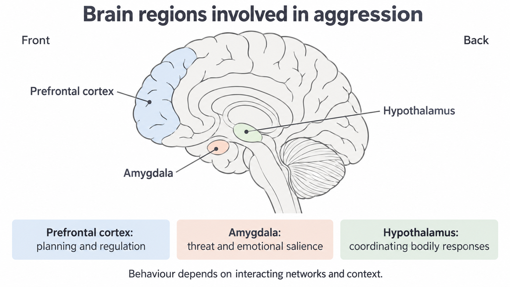
The picture is a simplified orientation guide, not a brain scan or an exact map of every connection. A claim about brain function needs evidence beyond where a structure appears in a drawing.
Explaining a response to provocation
A brain-based account of aggression considers how threat-related processing and the regulation of action work together.
Aggression is behaviour intended to harm another person who is motivated to avoid that harm. Anger is an emotional state; a person can feel anger without acting aggressively. Reactive aggression occurs in response to perceived provocation or threat. Proactive aggression is used deliberately to achieve a goal. An explanation of a rapid impulsive response should not automatically be applied to every planned aggressive act.
Imagine a fictional person who interprets a critical comment as a serious threat. Threat-sensitive processing involving the amygdala and connected regions can contribute to emotional arousal, while the hypothalamus helps coordinate bodily responses. Prefrontal networks can help the person consider the consequences and inhibit an inappropriate action. If regulation is impaired, acting on an immediate aggressive impulse may become more likely. This is an explanation of increased risk in a situation, not a claim that a damaged prefrontal cortex commands aggression.
The same comment can be interpreted differently because of earlier learning and the social context. A trusted friend's criticism may not be processed like a threat from someone who has previously been abusive. Brain functioning therefore provides a route through which environmental experiences can affect behaviour. Biological and environmental accounts can describe different levels of the same process.
This mechanism also gives a prediction: impairments affecting regulation could be associated with greater difficulty controlling reactive aggression. To test that prediction, researchers need measures of the relevant functioning and of behaviour. “Frontal damage” and “aggressive” are too broad on their own: the location, connections and behavioural measure matter.
What brain evidence can establish
Brain evidence supports a biological contribution most convincingly when the conclusion stays within the study's design and sample.
Raine et al. (1997) used positron emission tomography, or PET, to compare brain functioning during a continuous performance task in people charged with murder or manslaughter who pleaded not guilty by reason of insanity (NGRI), and matched controls. PET provided a measure related to glucose metabolism, rather than a direct reading of a person's intentions. The NGRI group showed reduced prefrontal glucose metabolism and other cortical and subcortical differences. This is consistent with the possibility of impaired regulatory functioning, but the detailed study is taught separately; its sample is not a sample of everybody who has been aggressive.
A strength is that a physiological measure gives evidence beyond participants' descriptions of how they feel. Finding a difference relevant to the proposed mechanism makes the explanation testable. The limitation is that a group comparison establishes an association. Researchers did not randomly create brain differences and then observe crime. Earlier experiences, injuries, substances or other characteristics could contribute to both functioning and behaviour. A difference during the task also does not show that the same pattern occurred during an offence.
Recent lesion-network research by Peng et al., published online in 2024 and in a 2025 journal issue, found that injuries associated with aggression could occur in different locations yet connect to a common network involving prefrontal regions. This supports investigating connections as well as isolated locations. It does not show that one network measurement can diagnose aggression in an individual, or that every connected structure has one function.
The evidence supports a qualified judgement: disrupted brain functioning can contribute to aggression risk, especially where a plausible regulatory mechanism is involved. It is insufficient to explain every aggressive act, identify a single aggression centre, or predict an individual from a group average. Biological explanations gain precision by specifying mechanisms; they lose explanatory power if they remove the person's learning and circumstances.
Brain injury and individual differences
A case study can reveal an important change after injury while still being an unsafe basis for predicting every patient's behaviour.
The historical case of Phineas Gage illustrates the connection between frontal injury and personality. He survived a workplace accident in 1848 in which a tamping iron passed through his head. His physician, John Harlow, later described changes in his behaviour, planning and social conduct. The case helped researchers consider frontal functioning in personality and regulation. It is not evidence that Gage became a murderer or that all frontal injuries produce aggression. The historical record lacks a modern standardised pre-injury personality assessment, and later reconstructions of the injury involve inference from surviving evidence.
Individual differences are ways people differ from one another, such as earlier personality, abilities, experience and circumstances. When a patient's behaviour changes, assuming there were no such differences before injury can exaggerate what the injury explains. A person who was already impulsive may differ from a person who was previously cautious. Their pre-injury history is needed to distinguish a new impairment from a longstanding characteristic.
Even a similar anatomical label can conceal different damage: injuries may differ in severity, side, exact region and connections affected. The age at injury also matters because the skills and circuits developing at that time differ. Plasticity, the capacity for neural organisation or functioning to change, can contribute to recovery, but it does not guarantee complete recovery or identical outcomes. Rehabilitation, support, stress, sleep and opportunities to practise regulation can affect how an impairment is expressed in daily life.
For a fictional pair of patients with broadly similar frontal injuries, stronger support and different earlier coping skills could help one manage difficult situations more effectively. This is a plausible explanatory illustration, not evidence from a conducted comparison. A careful real case study would combine medical evidence with pre-injury records, repeated behavioural observations and reports from relevant people. Multiple cases can show how consistent a pattern is, while preserving information about their differences.
An injury may contribute to a behavioural change. Its consequences still depend on the particular damage, the person, development and the environment.
This makes case evidence valuable for identifying possible mechanisms and challenging simple assumptions. Its rich individual detail is a strength, but one case cannot establish the proportion of all patients who will become aggressive. Labelling people as dangerous solely from their injury replaces a qualified scientific finding with an unsupported stereotype.