Biology 8.6 - 8.8 - Blood, vessels and the heart
Follow blood through its vessels and the four heart chambers, linking the structures of blood components, vessels and valves to transport and protection.
Blood: one tissue, four components
Blood is a tissue because it is a group of specialised components working together. Its liquid part, plasma, carries red blood cells, white blood cells and platelets through the circulatory system. The system transports useful substances to cells, removes waste substances and helps protect the body.
Red blood cells: carrying oxygen
Red blood cells are also called erythrocytes. Several features work together rather than one feature doing the whole job.
- A red blood cell is a biconcave disc: it is thinner in the centre than at the edge. This gives a large surface area compared with its volume and a short diffusion distance through the thin centre, so oxygen can enter or leave rapidly.
- A mature human red blood cell has no nucleus. This leaves more internal space for haemoglobin, the protein that binds oxygen.
- It is small and flexible, so it can bend through very narrow capillaries and take oxygen close to respiring cells.
The causal chain is therefore specialised shape and contents → efficient oxygen loading and unloading → oxygen transport from lungs to tissues. Red blood cells do not transport oxygen because they are red; the colour comes from the haemoglobin that performs the transport.
White blood cells: defending the body
White blood cells retain a nucleus and organelles, allowing them to carry out complex immune responses. Two groups are required here:
- Phagocytes can change shape. They surround, engulf and digest pathogens in a process called phagocytosis.
- Lymphocytes recognise foreign material. Some lymphocytes produce antibodies whose shapes bind specifically to antigens on a pathogen, helping the body target that pathogen.
White blood cells defend the body; they do not carry most of its oxygen. They are also much less numerous than red blood cells in healthy blood.
Plasma and platelets
Plasma is a pale liquid made mostly of water. Because many substances dissolve in water, plasma can transport dissolved glucose and other food molecules, carbon dioxide, urea, hormones and mineral ions. It also carries the blood cells and distributes thermal energy around the body. Most oxygen is carried by haemoglobin in red blood cells, not simply dissolved in plasma.
Platelets are small cell fragments rather than complete cells. At a damaged blood vessel they become sticky, gather at the wound and help a fibrin mesh form. The resulting clot reduces blood loss and forms a barrier to the entry of pathogens.
Higher tier: standard form and scale
A sample has red blood cells in . In , the estimate is cells. In standard form the first number is at least 1 and less than 10; multiplying the sampled volume multiplies the cell count in the same proportion. This standard-form calculation is Higher-tier mathematical content; the blood structure and function teaching applies to both tiers.
Blood vessels: built for pressure or exchange
A lumen is the hollow space through which blood flows. Arteries, veins and capillaries have different directions and conditions of flow, so their walls and lumens are different.
| Vessel | Direction and conditions | Structure → consequence → function |
|---|---|---|
| Artery | Carries blood away from the heart under relatively high, pulsing pressure | A thick wall containing muscle and elastic tissue withstands the pressure. Elastic tissue stretches as the heart pumps and recoils between beats, helping maintain flow. Its lumen is relatively narrow compared with the wall thickness. |
| Vein | Carries blood towards the heart under lower pressure | A thinner wall has less muscle and elastic tissue because it does not face arterial pressure. A wide lumen offers a low-resistance route. Pocket valves open towards the heart and close if blood starts to move backwards. |
| Capillary | Links the smallest arteries to the smallest veins and is the site of exchange with tissues | A wall only one cell thick gives a short diffusion distance. The narrow lumen brings blood close to the wall. Vast networks of capillaries provide a large total surface area and slow flow through the network, giving time for exchange. |
At body tissues, oxygen and dissolved food molecules move from capillary blood towards cells, while carbon dioxide and other wastes move towards the blood. The thin wall does not actively pull these substances across; it makes their diffusion route short.
Vessel names describe direction relative to the heart, not oxygen concentration. Most arteries in the body carry blood with a high oxygen concentration and most veins carry blood with a lower oxygen concentration, but the pulmonary artery and pulmonary vein are important exceptions.
An artery is not simply a vessel with oxygenated blood. It is a vessel that carries blood away from the heart; a vein carries blood towards the heart.
The heart and double circulation
The heart is a muscular organ that creates pressure to move blood. It has four chambers. The two upper atria receive blood; the two lower ventricles pump blood out. A muscular septum separates the right and left sides, preventing blood with lower oxygen concentration on the right from mixing with blood with higher oxygen concentration on the left.
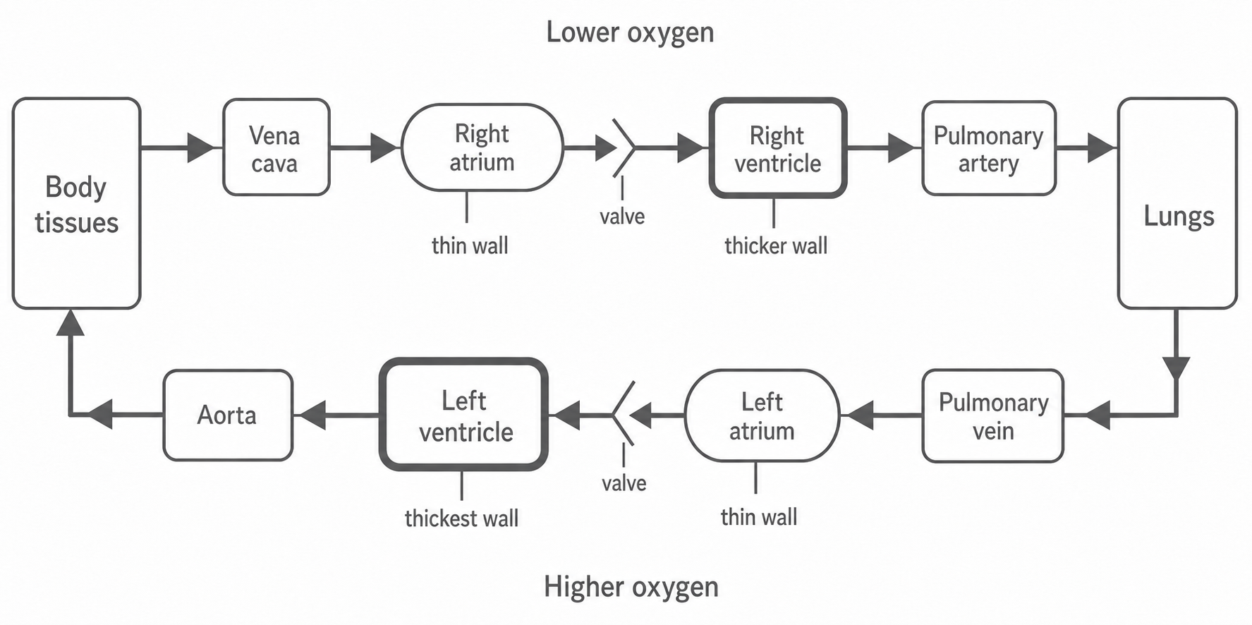
Follow one complete route:
- Blood returning from body tissues enters the right atrium through the vena cava. It has delivered much of its oxygen to cells.
- It passes through an atrioventricular valve into the right ventricle.
- The right ventricle contracts. Blood passes through an exit valve and enters the pulmonary artery, which carries it to the lungs.
- After gas exchange in the lungs, blood returns at a higher oxygen concentration through the pulmonary vein and enters the left atrium.
- It passes through another atrioventricular valve into the left ventricle.
- The left ventricle contracts. Blood passes through an exit valve into the aorta, which carries it to the body tissues.
This is a double circulatory system because blood passes through the heart twice in one complete journey: once through the pulmonary circuit between heart and lungs, and once through the systemic circuit between heart and the rest of the body.
[DIAGRAM: asset_slug: biology_8_6_8_10_blood_vessels_and_the_heart_diagram_01; description: Schematic double circulation, with correct vessel route, chamber wall thickness and atrioventricular valves. Exit valves are described in the text.]
Why the walls have different thicknesses
The chambers do not all perform the same amount of work.
- The atria have the thinnest walls because they push blood only a short distance into the ventricles.
- The right ventricle has a thicker wall than an atrium because it must pump blood to the lungs.
- The left ventricle has the thickest wall because it must produce higher pressure to send blood around the much longer systemic circuit.
Both ventricles move the same route of circulating blood in sequence; a thicker left wall does not mean the left ventricle should pump a different amount on each beat. It means that it generates greater pressure.
Why valves matter
Valves are flaps that respond to pressure differences. Forward pressure opens them; pressure beginning to act in the reverse direction closes them. They therefore prevent backflow and make contraction move blood along a one-way route. Valves guide flow but do not pump the blood themselves.
The diagram shows the route schematically rather than the anatomical positions of the chambers. It shows the valves between atria and ventricles; exit valves also lie between the ventricles and the aorta or pulmonary artery.