Biology 2.13 - Neurones and synapses
Trace electrical impulses through specialised neurones and chemical signals across synapses, then interpret reaction-time data as an example of nervous-system investigation.
Detecting and routing signals
A stimulus is a change in the internal or external environment. Heat, pressure, light and chemicals are examples. A sensory receptor is a specialised cell, group of cells or sensory nerve ending that detects a particular type of stimulus. Its structure makes it sensitive to that stimulus; when stimulated, it initiates an electrical impulse in a sensory neurone.
An electrical impulse is information carried along a neurone. It is not the neurone moving, and a neurone is not a hollow wire. The nervous system uses specialised cells to carry the message between locations:
- A sensory neurone carries an electrical impulse from a receptor towards the central nervous system.
- A relay neurone carries the impulse between neurones inside the central nervous system.
- A motor neurone carries an electrical impulse from the central nervous system to an effector.
The central nervous system (CNS) is the brain and spinal cord. An effector is a muscle or gland that produces a response; for example, a muscle contracts to move a limb. This gives a first route map:
stimulus → receptor → sensory neurone → CNS → motor neurone → effector → response
The route map does not yet show the relay neurone or the tiny junctions between neurones. Those become important when we zoom in.
Neurones built for transmission
A neurone is a specialised cell. Its long extensions carry an impulse over a distance, while branched regions allow it to receive or pass information at connections with other cells.
| Structure | Structure-function link |
|---|---|
| Cell body | Contains the nucleus and the cell machinery that keeps the neurone alive. Its position differs in sensory, relay and motor neurones. |
| Dendron | In the GCSE model of a sensory neurone, this is the long extension from the receptor towards the cell body. It carries an electrical impulse towards the cell body. |
| Axon | Carries an electrical impulse away from the cell body. In a sensory neurone it carries the impulse onwards into the CNS; in a motor neurone it carries the impulse from the CNS towards an effector. |
| Myelin sheath | Forms an electrically insulating covering around parts of a long nerve fibre. This insulation increases the speed at which an electrical impulse is transmitted. |
| Branched endings or dendrites | Provide regions where a neurone can receive information or connect with another cell. |
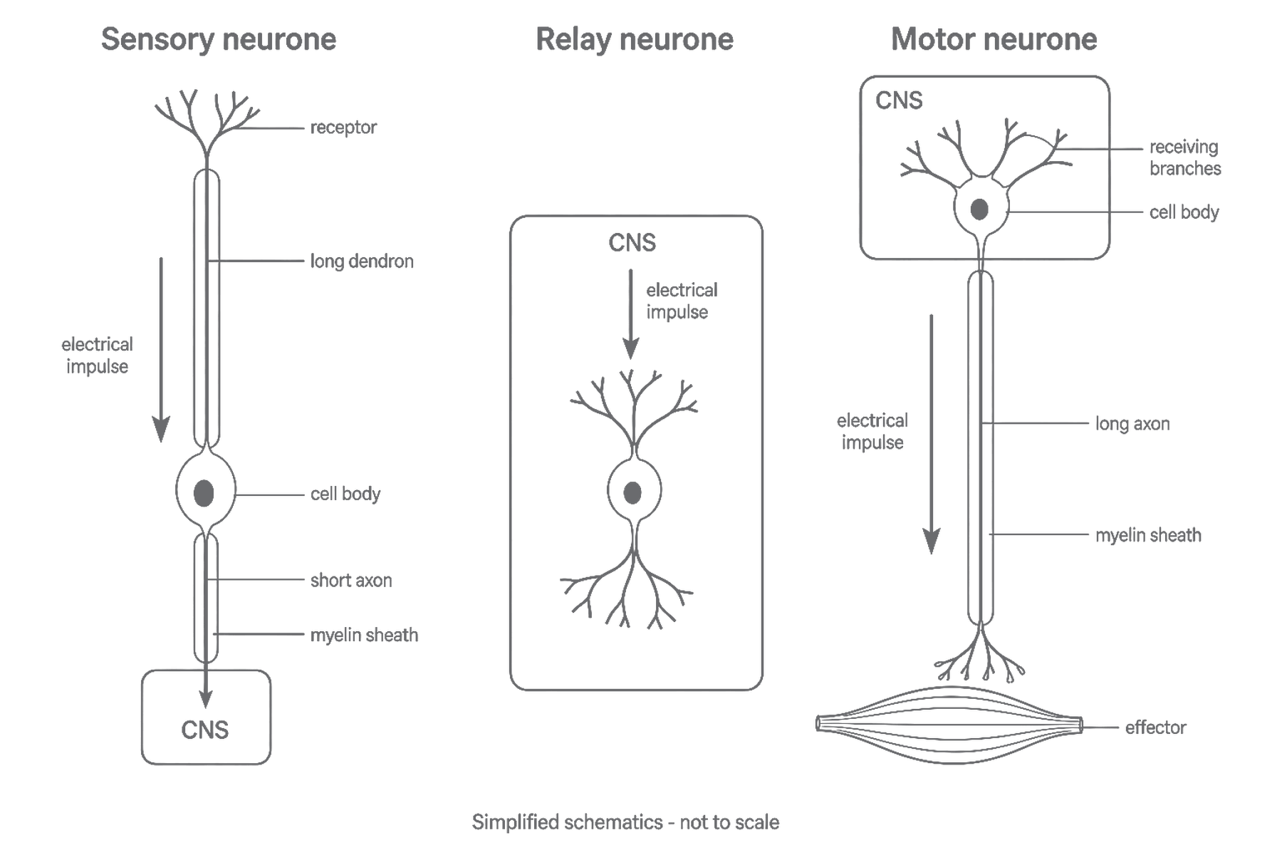
The three neurone types have structures suited to different positions in the route:
- A sensory neurone has a receptor ending, a long dendron leading towards a cell body positioned along the fibre, and an axon leading into the CNS. Myelin around its long fibre increases transmission speed.
- A relay neurone is short and branched because it connects neurones over short distances within the CNS. It receives an impulse from a sensory neurone and passes information towards a motor neurone.
- A motor neurone has its cell body and dendrites in the CNS and a long, often myelinated axon leading to an effector. Its branched ending passes the signal to a muscle or gland.
Notice that the name of a neurone tells you the direction and role of its signal, not simply what it looks like: sensory goes to the CNS, relay stays within the CNS, and motor goes from the CNS to an effector.
[DIAGRAM: asset_slug: bio_b_neurone_comparison; description: Three schematic neurones show dendron and axon direction, myelin, CNS location and an effector. Not to scale.]
Crossing a synapse
Neurones are separate cells. A synapse is the junction between one neurone and the next, including the tiny gap between them. The electrical impulse travels along the first neurone but does not simply jump across this gap as electricity.
Transmission across a synapse follows an ordered mechanism:
- An electrical impulse reaches the ending of the first, transmitting neurone.
- This causes the ending to release a chemical messenger called a neurotransmitter.
- Neurotransmitter molecules diffuse across the synaptic gap.
- They bind to matching receptor molecules on the membrane of the next neurone.
- This can trigger a new electrical impulse in the next neurone.
The signal is therefore electrical along a neurone, chemical across a synapse, then electrical along the next neurone. The neurotransmitter carries information across only the tiny gap; it does not flow along the whole reflex arc and it does not become the next electrical impulse.
The arrangement also gives transmission a direction: neurotransmitter is released from the ending of the transmitting neurone and acts on receptors on the receiving neurone. A synapse therefore passes the message from the first neurone to the next.
[DIAGRAM: asset_slug: bio_b_chemical_synapse; description: Electrical impulse arrives at a relay-neurone ending; neurotransmitter crosses a gap to receptors on the motor-neurone membrane and triggers a new impulse.]
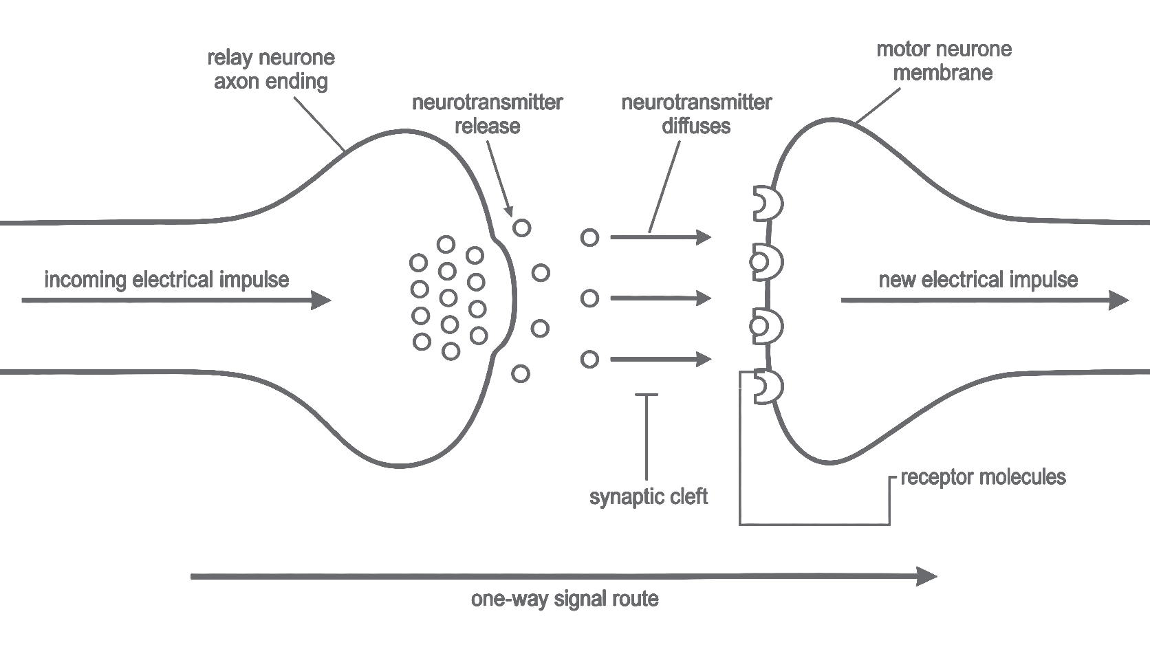
The figure shows one example, a relay-to-motor synapse. The same chemical-transfer idea applies at other synapses between neurones. A receptor molecule on a receiving neurone is different from a sensory receptor that detects a stimulus.
Reading a scatter diagram
A reaction-time task can provide data about a response involving the nervous system. In a simple computer task, a participant presses a key when a symbol appears. Sensory receptors detect the light, sensory neurones transmit information to the CNS, and motor neurones carry signals to the muscles. This is a voluntary reaction task, not a withdrawal reflex and not a direct measurement of the speed in one neurone.
Suppose a class investigates whether practice is associated with shorter reaction times. The fictional data are:
| Practice attempts | Reaction time / ms |
|---|---|
| 1 | 360 |
| 2 | 330 |
| 3 | 350 |
| 4 | 300 |
| 5 | 290 |
| 6 | 270 |
[DIAGRAM: asset_slug: bio_b_reaction_scatter; description: Portrait scatter diagram: six practice-attempt/reaction-time pairs, unjoined points, axes in attempts and ms; fictional data.]
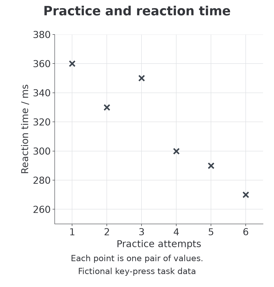
To translate the table into a scatter diagram, put practice attempts on the horizontal axis and reaction time, in milliseconds, on the vertical axis. Choose evenly spaced numerical scales, label both axes and plot one point for each pair. For example, the row becomes a point above 3 attempts and level with . Do not join successive dots: the purpose is to inspect the relationship between two variables.
The overall pattern slopes downwards: as the number of practice attempts increases, reaction time tends to decrease. This is a negative correlation. Smaller times mean faster responses. The third point is above the second, so the relationship is not perfect; use the overall pattern rather than demanding every pair follow it. A rising pattern would be a positive correlation, and scattered points with no trend would show no clear correlation.
This association does not prove that practice caused the change. Attention, fatigue and differences between participants could affect the readings. Keep the task and equipment the same, collect repeated measurements and compare appropriate groups before making a causal claim. A reaction time includes detection, CNS processing and muscle action, so it cannot be attributed to the axon alone.
Quick recap
Sensory neurones carry signals towards the CNS, relay neurones connect cells within it, and motor neurones carry signals to effectors. Myelin speeds transmission; neurotransmitters carry signals across synaptic gaps.