2.1.5(d)(i)-(ii) - Movement of molecules across membranes and model cells
Cells survive because their membranes are selective boundaries, not sealed walls. In this lesson you will learn how molecules move across membranes by diffusion, facilitated diffusion, active transport, endocytosis and exocytosis. You will then use model-cell investigations to connect those ideas to practical evidence, rates, graphs, uncertainty and surface area to volume ratio.
Choosing a transport route
A membrane transport question is usually solved by asking four linked questions.
- Is the substance moving down a concentration gradient or against it?
- Can it pass through the phospholipid bilayer directly, or does it need a membrane protein?
- Is ATP required as an immediate source of energy?
- Is the substance moving as individual molecules/ions, or is membrane vesicle movement involved?
[DIAGRAM: membrane_transport_routes: Lesson 043: Movement of molecules across membranes and model cells - diagram 01; asset_slug: 043_m02_1_5_movement_of_molecules_across_membranes_and_model_cells__diagram_01; recommended_method: drawn_biology; description: 16:9 deterministic drawn comparison of simple diffusion, facilitated diffusion, active transport, endocytosis and exocytosis with OCR-safe labels for gradient, protein use and ATP.]
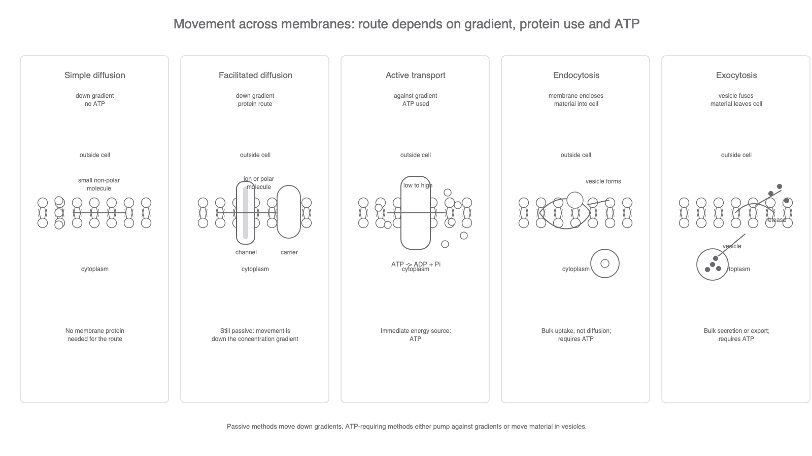
The main divide is passive versus ATP-requiring movement. Passive movement does not require ATP from the cell as an immediate energy source. Diffusion and facilitated diffusion are passive because the net movement is down a concentration gradient. Active transport, endocytosis and exocytosis require ATP.
| Route | Direction of net movement | Membrane protein needed? | ATP as immediate energy source? |
|---|---|---|---|
| Simple diffusion | Down a concentration gradient | No transport protein needed | No |
| Facilitated diffusion | Down a concentration gradient | Yes, channel or carrier protein | No |
| Active transport | Usually against a concentration gradient | Yes, carrier or pump protein | Yes |
| Endocytosis | Into the cell in vesicles | Membrane remodelling, not a channel | Yes |
| Exocytosis | Out of the cell in vesicles | Membrane fusion, not a channel | Yes |
The wording "net movement" matters. Molecules are always moving randomly, but if there are more molecules on one side of a membrane, more will move from that side to the other side per second. The overall result is movement from higher concentration to lower concentration until there is no concentration gradient. At that point, molecules still move in both directions, but there is no net movement.
Concentration gradient
A difference in the concentration of a substance between two regions.
A gradient is specific to one substance. A glucose gradient does not automatically tell you the direction of movement for ions, water or a different solute.
The quickest transport decision is: down-gradient without ATP is passive; protein-assisted but still down-gradient is facilitated diffusion; ATP-requiring pumping or vesicle movement is energy-dependent transport.
Diffusion and facilitated diffusion
Diffusion is the net movement of particles from a region where they are at a higher concentration to a region where they are at a lower concentration. In membranes, simple diffusion works best for small non-polar or lipid-soluble molecules because they can pass through the hydrophobic core of the phospholipid bilayer. Oxygen and carbon dioxide are standard examples.
Diffusion
The net movement of particles from a region of higher concentration to a region of lower concentration, down a concentration gradient, due to random movement.
Charged ions and many polar molecules cannot pass easily through the hydrophobic interior of the bilayer. They can still move passively if the membrane contains the correct transport protein. This is facilitated diffusion: the protein provides a route, but the concentration gradient still supplies the direction of net movement.
Facilitated diffusion
Passive movement of molecules or ions across a membrane through a specific membrane protein, down a concentration gradient.
Channel proteins form hydrophilic pores through the membrane. They are useful for ions and other small polar substances. Carrier proteins bind a particular molecule, change shape, and release the molecule on the other side. Both routes are selective, so a membrane can be permeable to one solute but not another.
Facilitated diffusion can reach a maximum rate because there is a limited number of channel or carrier proteins. Once the available proteins are working at full capacity, increasing the concentration gradient further may have little effect on rate. That plateau is not because ATP has run out; ATP was not required in the first place.
Do not write that facilitated diffusion is active transport just because a protein is involved. The key mark-scheme distinction is ATP use and movement relative to the concentration gradient.
Osmosis is water movement across a partially permeable membrane and has its own lesson next. For this lesson, treat it only as a boundary reminder: do not use "osmosis" as a general word for all membrane transport.
ATP-requiring movement
Active transport uses ATP as an immediate source of energy to move substances across membranes. It is usually taught as movement against a concentration gradient, from a lower concentration to a higher concentration. This cannot be explained by random movement alone, so a membrane carrier or pump protein changes shape using energy from ATP.
Active transport
Movement of molecules or ions across a membrane using ATP as an immediate source of energy, often against a concentration gradient.
The word "immediate" is important. Respiration makes ATP elsewhere in the cell, but active transport uses ATP directly at the membrane protein. A low ATP supply can therefore reduce active transport, even if the concentration gradient still favours the opposite passive movement.
Endocytosis and exocytosis also require ATP, but they are not the same as a pump moving one ion at a time. They move material using vesicles made from or fused with membranes.
| Process | Direction | What the membrane does | OCR-safe example idea |
|---|---|---|---|
| Endocytosis | Into the cell | Plasma membrane surrounds material and pinches off to form a vesicle | Uptake of large particles or fluid |
| Exocytosis | Out of the cell | Vesicle fuses with the plasma membrane and releases contents outside | Secretion of proteins or other cell products |
Both processes use membrane remodelling, so they can move substances that are too large for channels or carriers. They still need ATP because forming, moving and fusing vesicles requires energy.
Model cells for diffusion
A model cell is not a living cell. It is a practical system that represents one important feature of a cell so that the feature can be investigated under controlled conditions. For this lesson, the useful features are selective permeability, diffusion distance, concentration gradient, surface area and volume.
[DIAGRAM: model_cell_diffusion_rate_data: Lesson 043: Movement of molecules across membranes and model cells - diagram 02; asset_slug: 043_m02_1_5_movement_of_molecules_across_membranes_and_model_cells__diagram_02; recommended_method: drawn_biology; description: 16:9 deterministic practical and data visual showing a Visking tubing model cell, agar cube surface area to volume ratios and a diffusion-rate graph.]
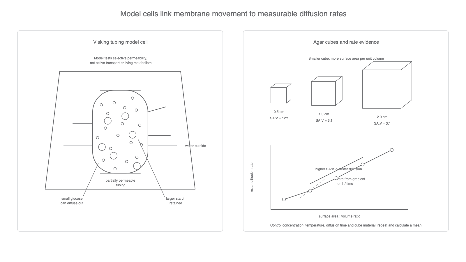
Visking tubing, also called dialysis tubing, is a common artificial membrane. It can be tied into a small bag, filled with a mixture such as glucose and starch solution, rinsed on the outside, and placed in water. After a set time, the water outside the tubing can be tested.
If glucose is detected outside but starch is not, the conclusion is not simply "diffusion happened". The stronger conclusion is that the membrane was permeable to the smaller glucose molecules but not to the larger starch molecules. The Visking tubing is a model for selective permeability, not a model for a complete plasma membrane.
To test the outside water, iodine solution can indicate starch if it turns blue-black, while Benedict's reagent, heated in a water bath, can indicate reducing sugar such as glucose by changing from blue towards green, yellow, orange or brick-red.
Agar cubes are another model. A cube containing indicator can be placed in acid, alkali or another diffusing solution. A colour change shows how far the solution has diffused into the cube after a fixed time, or how long it takes to reach the centre. Smaller cubes have a larger surface area to volume ratio, so more surface is available per unit volume for diffusion.
Rates, graphs and surface area
Diffusion investigations need a measured rate, not just a statement that a colour changed. The rate may be measured as:
- distance diffused per unit time
- mass or volume change per unit time
- concentration change per unit time
1 / timewhen the endpoint is the time taken for a fixed change
Diffusion rate
For a cube, surface area and volume can be calculated from its side length. This is useful because diffusion across the outside depends on surface area, while the amount of material supplied by diffusion depends on volume.
Surface area to volume ratio and rate
A cube of agar has side length 1.0 cm.
Surface area is measured in square units, so the unit is cm2, not cm3.
If the colour change reaches the centre in 240 s, a relative rate can be calculated as:
The biological interpretation is that a higher surface area to volume ratio gives a faster diffusion rate into the whole model cell.
When data are plotted, put the independent variable on the x-axis and the dependent variable on the y-axis. For example, surface area to volume ratio belongs on the x-axis if it is the factor being changed, and mean diffusion rate belongs on the y-axis if it is the measured outcome.
If the graph is a straight line, rate of change can be found from the gradient:
Gradient
If the graph is curved and you need the rate at one point, use the gradient of a tangent at that point. This is especially useful when the initial diffusion rate is faster than later diffusion, because the concentration gradient becomes smaller as diffusion proceeds.
Uncertainty should shape the conclusion. A stopwatch result may include reaction-time uncertainty. A ruler measurement may include uncertainty from the smallest division. Repeats and a mean improve reliability, but they do not fix an invalid method where two variables were changed at the same time.
Designing and evaluating model-cell investigations
A valid PAG8-style model-cell diffusion investigation changes one independent variable and controls the others. Depending on the method, the independent variable could be concentration gradient, cube size, diffusion distance, surface area to volume ratio, temperature, or molecule size. The dependent variable must be measurable, such as time for colour change, distance diffused, concentration of glucose outside the tubing, mass change, or absorbance.
Controlled variables should match the factor being tested. In a Visking tubing investigation, useful controls include tubing length, volume inside the tubing, outside volume, starting concentration, time, temperature and how thoroughly the outside of the tubing is rinsed. In an agar cube investigation, useful controls include agar composition, indicator concentration, solution concentration, solution volume, temperature and time in solution.
Reliability comes from repeats, means and checking anomalies. Validity comes from whether the method really tests the intended factor. For example, comparing two agar cubes of different size can test surface area to volume ratio only if the agar composition and external solution are the same. Otherwise, cube size is not the only explanation for any change in rate.
Model limitations should be stated precisely.
| Model | Useful because | Limitation |
|---|---|---|
| Visking tubing | Shows selective permeability and diffusion of small molecules | Not a phospholipid bilayer; no membrane proteins, ATP use or metabolism |
| Agar cube | Shows diffusion distance and surface area to volume ratio | Not a living cell; diffusion happens through gel, not a real plasma membrane |
When evaluating a model, do not just say "it is not a real cell". State the exact feature it models well and the exact feature it cannot model.
Use that precision in the final self-explanation: name the route first, then justify it from gradient, protein use, ATP or vesicle movement.