2.1.5(a)-(b) - Membrane roles and the fluid mosaic model
Membranes are not just the edges of cells. They are working surfaces that separate aqueous spaces, organise reactions and allow cells to receive signals. In this lesson you will learn the OCR roles of membranes and then use the fluid mosaic model to explain how the named membrane components make those roles possible.
Why Membranes Matter
A biological membrane is a thin structure built mainly from a phospholipid bilayer with other molecules embedded in or attached to it. The cell-surface membrane, also called the plasma membrane, surrounds the cell. Eukaryotic cells also have membranes around organelles and membranes inside some organelles.
Partially permeable membrane
A partially permeable membrane allows some substances to cross more easily than others, so it can separate two regions while still allowing controlled exchange.
The first OCR role is as a partially permeable barrier. At the surface of a cell, the membrane separates the cytoplasm from the external environment. Around an organelle, a membrane separates the organelle contents from the cytoplasm. Within organelles, folded or stacked membranes can create separate reaction surfaces and spaces.
This barrier role is not the same as being impermeable. If a membrane were completely impermeable, cells could not take in required substances, remove waste products or respond to their surroundings. The key idea is controlled separation: the membrane keeps regions distinct while making selective movement possible. The exact movement mechanisms come in later lessons.
The second role is as a site of chemical reactions. Many reactions happen at membrane surfaces because enzymes and other proteins can be held in place there. For example, membranes inside mitochondria and chloroplasts provide organised surfaces for later energy-transfer processes; you do not need the details of those pathways in this lesson.
The third role is cell communication. A cell can detect chemical signals when molecules bind to membrane components on its surface. This is why membranes matter to coordination in a multicellular organism and to the action of some drugs.
Fluid Mosaic Model
The fluid mosaic model is the OCR model for membrane structure. It explains the membrane as a phospholipid bilayer with a mixture of other components, including proteins, glycoproteins, glycolipids and cholesterol.
The word "fluid" means the membrane is not a rigid sheet. Phospholipids and many membrane proteins can move sideways within the plane of the membrane. This gives the membrane flexibility and allows components to be arranged for different functions.
The word "mosaic" means the membrane contains a varied pattern of molecules. A membrane is not made from phospholipids alone. Proteins may span the bilayer or sit at one surface, carbohydrate chains may be attached to proteins or lipids, and cholesterol may sit among the phospholipid tails.
[DIAGRAM: fluid_mosaic_membrane_model: Lesson 041: Membrane roles and the fluid mosaic model - diagram 01; asset_slug: 041_m02_1_5_membrane_roles_and_the_fluid_mosaic_model__diagram_01; recommended_method: drawn_biology; description: A clean 16:9 drawn biology schematic of the fluid mosaic model showing phospholipid bilayer, hydrophilic heads, hydrophobic tails, cholesterol, membrane protein channel, glycolipid, glycoprotein receptor, hormone/drug binding, hydrophobic core and the OCR roles of membranes.]
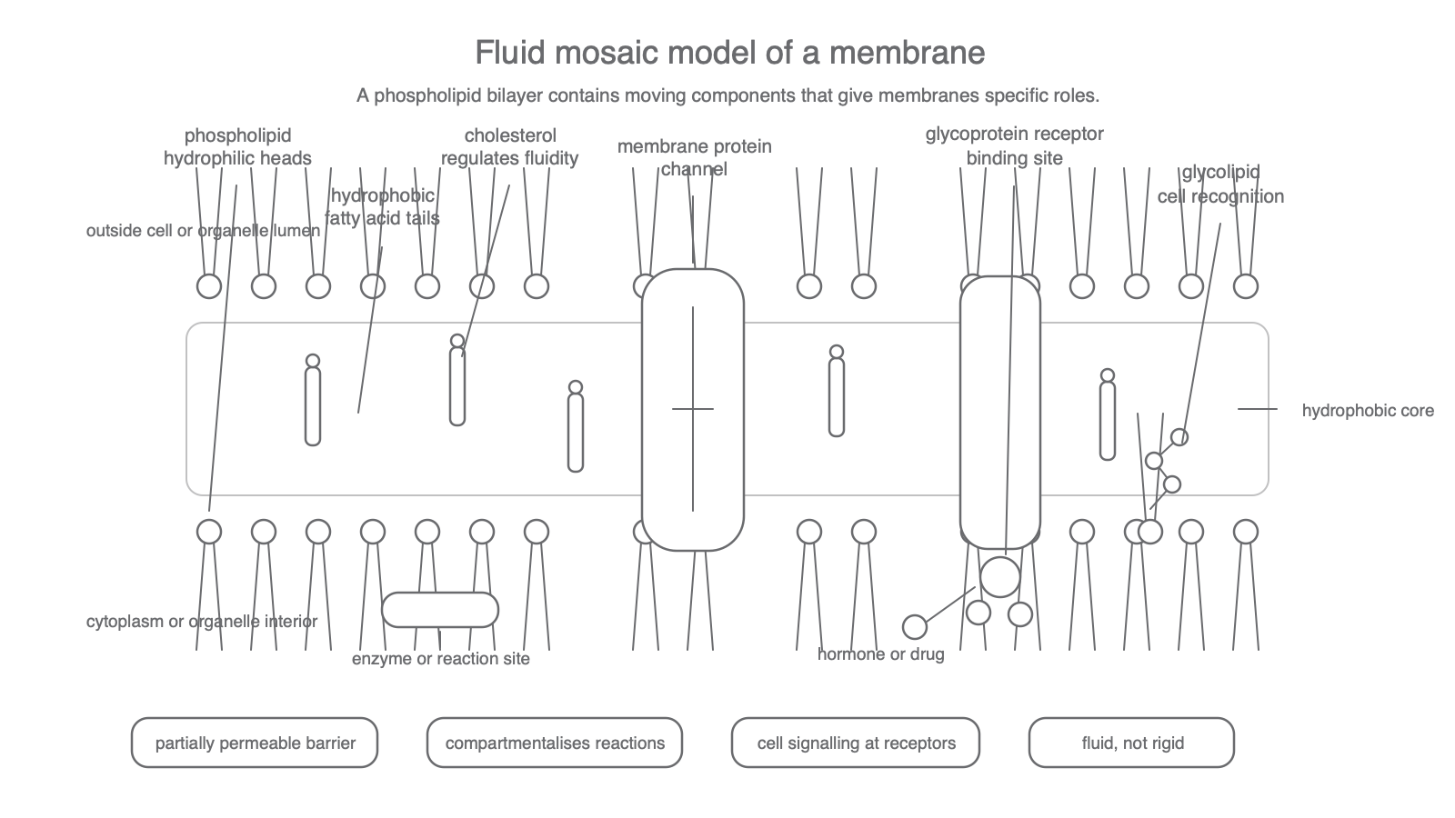
Read the diagram as a model, not as a claim that every membrane has exactly the same proportions of components. The phospholipid bilayer gives the basic barrier. The embedded or attached components give more specific functions such as recognition, enzyme activity, signalling and regulated movement. Transport mechanisms are a later lesson; here the important point is that membrane proteins are part of the mosaic.
Component-Function Links
Strong OCR answers link each component to its role. A list of names is rarely enough; the mark often comes from explaining what the component does in the membrane.
| Component | Position or structure | OCR-safe role |
|---|---|---|
| Phospholipids | Hydrophilic heads face aqueous regions; hydrophobic tails point inwards | Form the bilayer and hydrophobic core, giving a partially permeable barrier |
| Cholesterol | Fits among phospholipid tails in many animal cell membranes | Regulates membrane fluidity and helps control permeability |
| Membrane proteins | Embedded in, spanning or attached to the bilayer | Can act as channels, carriers, enzymes or receptors, depending on the protein |
| Glycolipids | Lipids with carbohydrate chains attached | Help cell recognition and membrane stability |
| Glycoproteins | Proteins with carbohydrate chains attached | Help cell recognition and may act as receptors |
Phospholipids are amphipathic: each molecule has a hydrophilic head and hydrophobic tails. In water, the hydrophilic heads face the aqueous solution outside the cell or in the cytoplasm, while the hydrophobic tails point away from water. This creates a bilayer with a hydrophobic core. That core helps explain why the membrane is partially permeable rather than a free opening between two solutions.
Cholesterol should be described carefully. It is a membrane component required by OCR, and in animal cell membranes it helps regulate fluidity and permeability. It is not a separate layer, and it is not the same as a phospholipid. A safe answer is: cholesterol fits between phospholipid tails and helps stabilise membrane fluidity.
Proteins are the most varied membrane components. Some are intrinsic proteins, meaning they are embedded in the bilayer; a channel protein is one example. Other proteins are associated with one surface. Proteins can act as enzymes, receptors, channels or carriers. In this lesson, you only need the component-function idea, not the full mechanisms of transport across membranes.
Glycolipids and glycoproteins both involve carbohydrate chains projecting from the membrane. The difference is the molecule they are attached to: a glycolipid is carbohydrate attached to a lipid, while a glycoprotein is carbohydrate attached to a protein. These carbohydrate chains are useful for recognition between cells and for receptors at the cell surface.
Receptors And Signalling
Cell signalling depends on a signal being detected by a target cell. A membrane-bound receptor is a protein, often a glycoprotein, with a binding site for a particular molecule.
Membrane-bound receptor
A membrane-bound receptor is a membrane protein with a specific binding site where a signalling molecule, such as a hormone, or a drug can bind.
Hormones are chemical messengers. A hormone can be carried around the body, but only cells with a complementary receptor respond strongly to it. Binding to the receptor can start a response inside the target cell. You do not need the full signalling pathway here; the required idea is receptor binding at the membrane.
Some drugs act in the same general way. A drug molecule may bind to a membrane-bound receptor and change the activity of the target cell. This does not mean the drug must always enter the cell. It may act by binding to a receptor at the cell-surface membrane.
This is a useful place to avoid a common misconception: a receptor is not simply a hole in the membrane. A receptor is a specific binding site, usually on a protein. A channel may allow movement of substances across the membrane, but receptor binding is about detecting a signal.
Model, Scale And Precision
The fluid mosaic model is a scientific model. It simplifies a very small, dynamic structure so that it can explain several observations at once: membranes are flexible, they separate aqueous regions, they contain proteins and they allow specific communication through receptors.
When asked to describe the fluid mosaic model, include both parts of the phrase: "fluid" means components can move within the membrane, and "mosaic" means different components are scattered through the bilayer.
OCR flags M0.2 for this row, so you may need to recognise standard form in a membrane context. A plasma membrane is often only a few nanometres thick. Since 1 nm is 1 x 10^-9 m, a membrane thickness of 7.5 nm is:
Converting membrane thickness
7.5 nm = 7.5 x 10^-9 m
The number 7.5 stays the same and the unit changes from nanometres to metres using the factor 10^-9.
The biological interpretation is more important than the number itself: membranes are far too thin to resolve as detailed structures with a light microscope, so the model is an explanation of molecular organisation, not something you would see directly in an ordinary school light microscope.
Keep the boundary clean. Temperature, solvents, beetroot pigment release, diffusion, active transport and osmosis are all important membrane topics, but they belong to later lessons in this sequence. Here, the required content is roles, model and named components.
Explain It Back
Use this final diagnostic after you have worked through the model and the component roles. It is not asking for new detail; it is checking whether the structure-function links are clear.