2.1.2(s)(i)-(ii) - Paper and thin layer chromatography
In this lesson you are learning how paper chromatography and thin layer chromatography separate biological compounds, how to run a valid practical investigation, and how to calculate and interpret Rf values. OCR cares about the method because a chromatogram is evidence: it can show whether a biological solution contains more than one compound and whether an unknown behaves like a known standard. The tight boundary here is paper and TLC chromatography of biological molecules such as proteins, carbohydrates, vitamins and nucleic acids.
Separation Principle
Chromatography separates compounds in a mixture because the compounds do not all move with the solvent to the same extent. Each compound spends some time associated with a stationary phase and some time dissolved in a mobile phase.
Stationary phase
The stationary phase is the material that does not move during chromatography, such as chromatography paper or the thin solid layer on a TLC plate.
Mobile phase
The mobile phase is the solvent that moves through or over the stationary phase and carries dissolved compounds with it.
In paper chromatography, the stationary phase is the paper. In thin layer chromatography, or TLC, the stationary phase is a thin layer of solid material such as silica gel, alumina or cellulose on a support. In both methods, the mobile phase is a solvent or solvent mixture that rises by capillary action.
The key idea is relative attraction. A compound that is more soluble in the mobile phase, or less strongly attracted to the stationary phase, moves further. A compound that is less soluble in the mobile phase, or more strongly attracted to the stationary phase, moves a shorter distance. This is why one biological solution can separate into several spots.
Paper chromatography and TLC can be used to analyse biological solutions containing compounds such as amino acids from proteins, sugars or other carbohydrates, vitamins, and nucleotides or nucleic-acid-related compounds. The lesson is not asking you to learn all the chemistry of those molecules here; it is asking you to apply the same separation principle to biological compounds.
[DIAGRAM: chromatogram-rf-measurement: Lesson 029: Paper And Thin Layer Chromatography - diagram 01; asset_slug: 029_m02_1_2_paper_and_thin_layer_chromatography__diagram_01; recommended_method: drawn_biology; description: Developed paper or TLC chromatogram showing the baseline, solvent front, separated spots, measurement to the centre of a spot, measurement to the solvent front, and the Rf formula.]
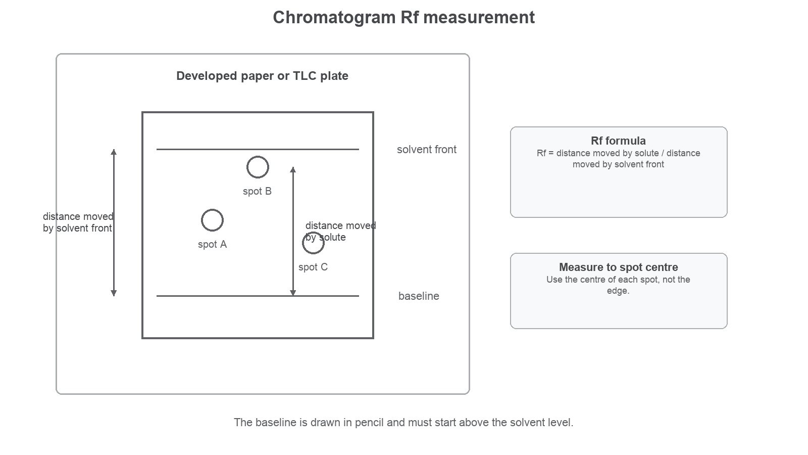
If you explain why one spot moved further, name the phases. "It is more soluble" is incomplete; "it is more soluble in the mobile phase" or "it is less attracted to the stationary phase" is the creditworthy idea.
Valid Chromatography Method
A good chromatogram starts with careful setup. Draw the baseline in pencil near the bottom of the paper or TLC plate. Pencil is used because it does not dissolve and move with the solvent in the same way as many inks.
Place a small, concentrated sample spot on the baseline using a capillary tube or suitable applicator. If the spot needs to be more concentrated, let it dry and then add another small spot exactly on top. A large wet spot spreads sideways and can produce smearing, so the separated spots become difficult to measure or compare.
Put the paper or TLC plate into a chromatography tank with the solvent level below the baseline. If the solvent covers the baseline, the sample dissolves directly into the solvent reservoir rather than being carried up through the stationary phase.
As the solvent rises, it carries some compounds further than others. Remove the paper or plate before the solvent front reaches the top, then mark the solvent front immediately in pencil. The solvent front is the furthest point reached by the solvent. It can fade as the solvent evaporates, so failing to mark it can make the Rf calculation invalid.
The usual method sequence is:
| Method step | Why it matters |
|---|---|
| Draw a pencil baseline | Gives a fixed origin that will not dissolve into the solvent |
| Apply a small dry concentrated spot | Reduces smearing and improves separation |
| Keep the solvent below the baseline | Prevents the sample washing into the solvent reservoir |
| Cover the tank if instructed | Reduces solvent evaporation and helps keep conditions consistent |
| Remove the plate before the solvent reaches the top | Leaves a measurable solvent front |
| Mark the solvent front immediately | Preserves the distance needed for Rf calculation |
| Run known standards alongside unknowns | Allows comparison under the same conditions |
Risk management is part of the method. Some extraction and running solvents are hazardous or flammable, so a practical may need good ventilation, a fume cupboard, eye protection and correct solvent disposal. If a TLC plate is viewed under ultraviolet light, the UV source must be used safely. Handle paper or TLC plates carefully and avoid touching the surface, because oils from fingers can contaminate the stationary phase and alter spot movement.
Rf Values
An Rf value is a ratio that describes how far a solute moved compared with the solvent front. It lets you compare spots from a chromatogram using numbers rather than only descriptions.
Rf Value
Both distances are measured from the same baseline. For a spot, measure from the baseline to the centre of the spot. For the solvent, measure from the baseline to the solvent front. Rf has no unit because a distance divided by a distance cancels the units.
Calculating Rf
A spot moved 42 mm from the baseline. The solvent front moved 80 mm from the same baseline.
Use the formula:
Substitute the values:
The distances were measured to two significant figures, so a sensible reported value is:
The value has no unit. Biologically, this spot moved just over half as far as the solvent front under these chromatography conditions.
You may also need to rearrange the equation. If you know the Rf value and the distance moved by the solvent, then:
Distance Moved By Solute
Rearranging Rf
A known vitamin has an Rf value of 0.62 in a particular solvent. The solvent front moved 75 mm.
To an appropriate whole-millimetre measurement, the expected spot centre would be about 47 mm from the baseline.
Interpreting Chromatograms
A chromatogram is strongest when an unknown is run alongside known standards. A standard is a known compound treated under the same chromatography conditions. If an unknown spot has the same Rf value as a standard, that supports the idea that the unknown contains that compound.
The phrase "under the same conditions" matters. Rf depends on the solvent, stationary phase, temperature, thickness of the TLC layer, and technique. An Rf value from one solvent system should not be compared as if it were universal.
Imagine an unknown biological solution gives three spots after TLC. The solvent front moved 80 mm.
| Spot | Distance from baseline to spot centre / mm | Rf |
|---|---|---|
| Known carbohydrate X | 24 | 0.30 |
| Known vitamin Y | 44 | 0.55 |
| Unknown spot 1 | 24 | 0.30 |
| Unknown spot 2 | 44 | 0.55 |
| Unknown spot 3 | 62 | 0.78 |
The unknown contains at least three detectable compounds because it produced three spots. Spot 1 has the same Rf as carbohydrate X under these conditions, so the chromatogram supports the conclusion that carbohydrate X is present. Spot 2 supports the conclusion that vitamin Y is present. Spot 3 does not match either standard in this run, so it should be described as an unidentified compound rather than guessed.
Use careful conclusion language. "The unknown may contain X because the spot has the same Rf as X under the same conditions" is stronger than "the unknown is definitely X". A different compound could sometimes have the same Rf in one solvent system.
Rf can also support explanation. If spot 3 has the highest Rf, it moved furthest relative to the solvent front. That suggests it was more soluble in the mobile phase, or less strongly attracted to the stationary phase, than the other spots under these conditions.
Do not use spot size alone as a precise measure of concentration in this lesson. A darker or larger spot may suggest more material was present or loaded, but spot size can also be affected by spotting technique, visualisation and smearing. OCR expects you to be cautious about what the evidence actually shows.
Practical Investigation Evaluation
Paper chromatography and TLC are often used as practical investigations rather than just demonstrations. The practical question might be: does this solution contain more than one amino acid, sugar derivative, vitamin or nucleic-acid-related compound? Is an unknown compound consistent with a known standard? Which solvent system gives the clearest separation?
To make that evidence valid, control the variables that affect movement:
| Variable or decision | Why it affects validity |
|---|---|
| Same solvent system | Changes solubility in the mobile phase and therefore Rf |
| Same stationary phase | Changes attraction to the stationary phase |
| Same baseline and development distance | Makes spot and solvent distances comparable |
| Similar spot size and concentration | Reduces smearing and unfair differences in visibility |
| Standards run on the same plate or paper | Allows direct comparison under identical conditions |
| Plate not touching the tank side | Prevents uneven solvent movement or condensation effects |
| Solvent front marked immediately | Preserves the distance for Rf calculation |
When evaluating a method, link the improvement to the evidence. "Repeat the chromatography" is useful if it checks repeatability. "Use known standards" is useful because the unknown can be compared with compounds treated under the same conditions. "Use a different solvent" is useful if the original solvent did not separate two compounds clearly.
Improving Separation
A paper chromatogram of a nucleic-acid-related solution shows two spots so close together that their centres are difficult to measure. A useful improvement is to test a different solvent system or a longer development distance, provided the solvent front still remains on the paper. This may give better separation, making Rf values easier to measure and reducing uncertainty in identification.
A common misconception is that a high Rf means a compound is more attracted to the stationary phase. It is the opposite for paper/TLC under the same run conditions. A high Rf means the compound moved further with the mobile phase, so it was more soluble in the mobile phase or less strongly attracted to the stationary phase.
Practical Investigation Evaluation Continued
To check your understanding, explain the whole method to yourself in plain language as if teaching a younger student: "A mixture starts on a baseline, a solvent moves through a fixed material, and each compound moves a different distance." Then add one limitation to the explanation: an Rf match supports identification only when the unknown and standard are run under the same conditions.
Paper and TLC chromatography turn a biological mixture into evidence by separating compounds, measuring spot movement against the solvent front, and comparing Rf values carefully under matched conditions.
Quick Check
Use this as a short comprehension check on the section above.