2.1.1(g)-(h) - Eukaryotic cell ultrastructure and photomicrographs
This lesson builds a usable map of eukaryotic cell components and then shows how to interpret electron micrographs without guessing. The key habit is structure-function reasoning: identify what is visible, then connect it to a precise function.
Ultrastructure Meaning
Ultrastructure means cell detail that is usually seen with an electron microscope rather than a light microscope. It includes small organelles and membrane systems that cannot be resolved clearly with ordinary light microscopy.
Eukaryotic cells contain membrane-bound organelles. Plant and animal cells share many components, but plants also have structures such as a cellulose cell wall and chloroplasts. Some structures, such as ribosomes, are not membrane-bound but are still part of the required eukaryotic cell content.
[DIAGRAM: eukaryotic_ultrastructure_map: Lesson 015: Eukaryotic ultrastructure OCR components - diagram 01; asset_slug: 015_m02_1_1_eukaryotic_cell_ultrastructure_and_photomicrographs__diagram_01; recommended_method: drawn_biology; description: Deterministic labelled map of OCR-required plant and animal eukaryotic cell components.]
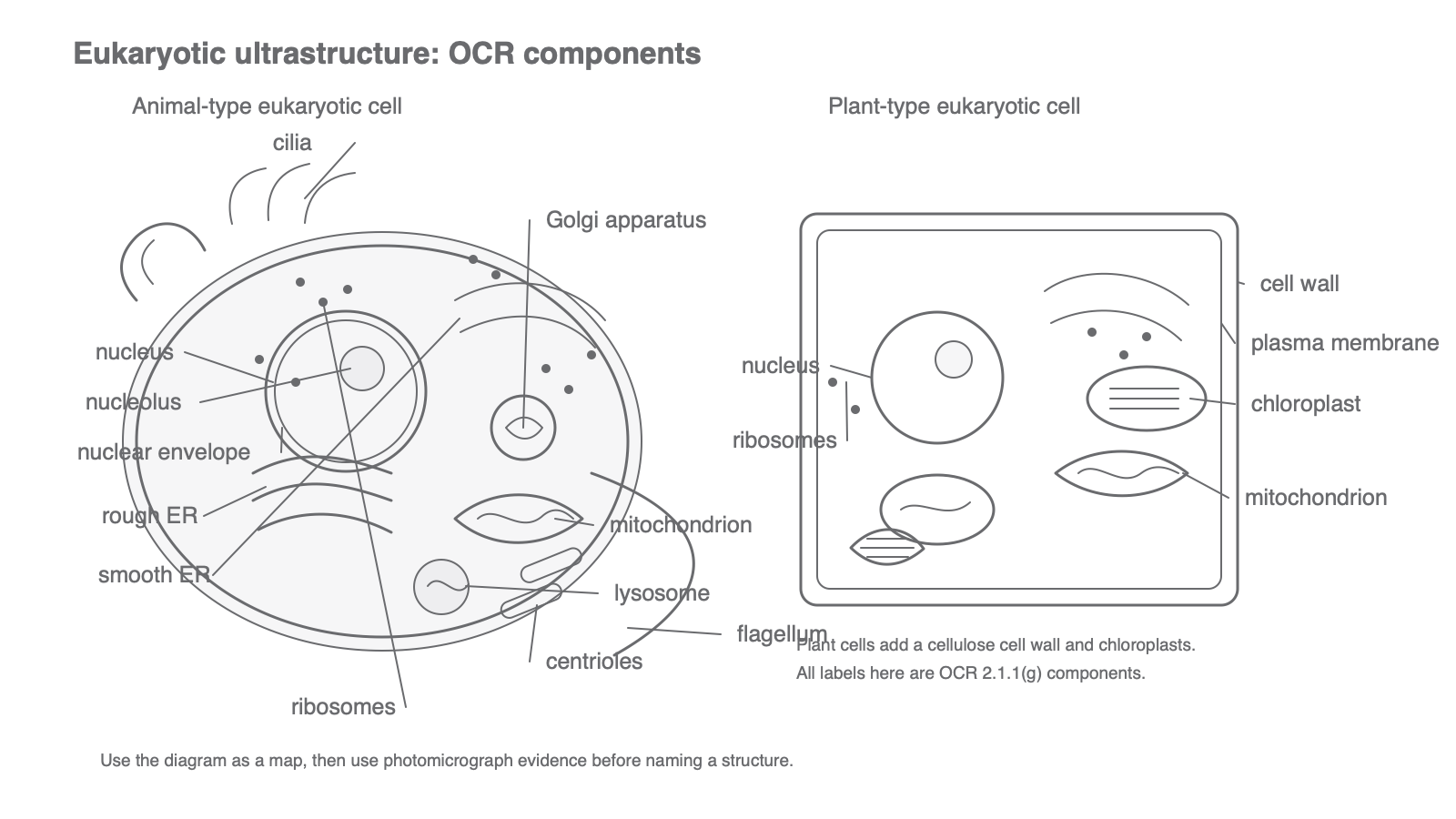
Organelle Functions
Learn each component by asking what job it makes possible.
| Component | Outline function |
|---|---|
| Nucleus | contains DNA and controls gene expression/cell activities |
| Nucleolus | makes ribosomal RNA and assembles ribosome subunits |
| Nuclear envelope | double membrane around nucleus with pores for exchange |
| Rough ER | folds and transports proteins made on attached ribosomes |
| Smooth ER | makes lipids and helps with detoxification in some cells |
| Golgi apparatus | modifies, sorts and packages proteins or lipids into vesicles |
| Ribosomes | site of protein synthesis; not membrane-bound |
| Mitochondria | site of aerobic respiration and ATP production |
| Lysosomes | contain digestive enzymes for breakdown inside the cell |
| Chloroplasts | site of photosynthesis in plant cells |
| Plasma membrane | controls movement into and out of the cell |
| Centrioles | involved in spindle organisation during cell division in animal cells |
| Cell wall | cellulose support and shape in plant cells |
| Flagella | longer projections that can move a cell |
| Cilia | shorter projections that can move fluid or material over a cell surface |
For a one-mark organelle identification, give one clear name. Two contradictory names usually lose the precision that mark schemes expect.
Membranes And Non-Membranes
Many eukaryotic organelles are membrane-bound, including the nucleus, ER, Golgi apparatus, mitochondria, lysosomes and chloroplasts. Membranes separate reactions into compartments and help control conditions inside each organelle.
Ribosomes are not membrane-bound. A common error is to call every visible dot in an electron micrograph a vesicle or a lysosome. Ribosomes are small dense particles and may appear free in the cytoplasm or attached to rough ER.
The plasma membrane is not just a boundary line. It controls movement into and out of the cell. The plant cell wall lies outside the plasma membrane and gives support; it is not the membrane controlling exchange.
Interpreting Photomicrographs
Photomicrographs are microscope images. In this lesson, the important OCR skill is interpreting TEM and SEM images.
[DIAGRAM: photomicrograph_interpretation_cues: Lesson 015: Photomicrograph cues for OCR electron images - diagram 02; asset_slug: 015_m02_1_1_eukaryotic_cell_ultrastructure_and_photomicrographs__diagram_02; recommended_method: drawn_biology; description: Deterministic TEM and SEM cue sheet showing internal section evidence, surface evidence, scale and cautious interpretation.]
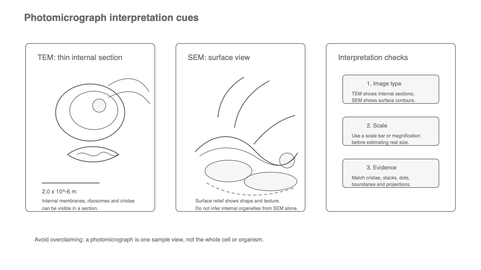
TEM images show internal structure in thin sections. A mitochondrion often appears as an oval or elongated structure with internal folds called cristae. A lysosome is more likely to be a small vesicle-like body without those internal folds. This distinction matters because students often confuse them.
SEM images show surface features. They may show surface projections such as cilia, but they do not show the internal arrangement of organelles.
Electron micrographs are prepared evidence. Sections may cut through only part of an organelle, false colour may be added, and absence from one image is not proof that the cell lacks a structure.
Scale And Standard Form
Cell images often use very small measurements. Exact organelle sizes are not a recall requirement here, but you must be comfortable reading ordinary and standard form values when a question supplies them.
For example:
because one micrometre is metres.
Using A Scale Bar
A TEM scale bar is labelled . A mitochondrion is three scale-bar lengths long.
Actual length:
This is 3.0 micrometres.
Using Evidence Not Memory
The safest approach to a photomicrograph is evidence first, name second:
- Is it a TEM internal section or an SEM surface view?
- What visible features are present?
- Which organelle/component fits those features?
- What function follows from that component?
Do not add structures that are not visible. Do not assume every plant cell image shows chloroplasts, or every animal cell image shows centrioles. The question can only reward what the evidence supports.
Using Evidence Not Memory Summary
Eukaryotic ultrastructure is a set of named components plus functions; photomicrograph interpretation is the disciplined use of visible evidence.