2.1.1(a) - Microscopy to observe eukaryotic cells
This lesson is about using microscopy as evidence: choosing a microscope because of the kind of cell structure you need to investigate, then interpreting what its image can and cannot show. The focus is eukaryotic organisms, so examples may come from animal, plant, fungal or protist cells. You are not yet doing the detailed slide preparation, staining, drawing or magnification calculations from the neighbouring OCR rows.
Microscopy As Evidence
Microscopy means using a microscope to observe structures that are too small to see clearly with the unaided eye. In Biology, that matters because cells are the basic units of living organisms, but many of their structures are below the scale of ordinary vision.
Eukaryotic cell
A cell with genetic material enclosed in a nucleus and with membrane-bound organelles in the cytoplasm.
Eukaryotic organisms include animals, plants, fungi and protists. The exact cell type changes, but the purpose of microscopy stays the same: it lets a biologist connect a visible image with a question about cell structure. For example, a biologist might ask whether plant cells in a leaf contain visible chloroplasts, whether an animal tissue contains differently shaped cells, or whether a cell surface has projections.
The microscope does not simply make "the truth" appear. It produces an image using a particular method. A useful interpretation therefore depends on matching the image to the microscope that made it.
| Question being investigated | What the image needs to show |
|---|---|
| Are whole cells present and arranged in a tissue? | Overall cell shape, tissue layout or larger cell structures. |
| What internal ultrastructure is present inside a cell? | Thin-section internal detail, such as organelle membranes. |
| What does the surface of a cell or tissue look like? | Surface shape, projections and three-dimensional-looking topography. |
Microscopy is useful because different microscopes provide different kinds of cell evidence, not because one microscope is automatically "best" for every biological question.
Light Microscope Images
A light microscope uses visible light and glass lenses to form an image. In a school or college setting, it is often the first microscope used because it can show whole cells and larger cell structures without needing the complex preparation required for electron microscopy.
Light microscope images can show features such as cell outlines, nuclei in stained animal cells, chloroplasts in some plant cells, and the arrangement of cells in a thin tissue section. They can also be used with living or fresh specimens in suitable preparations, which is useful when a biologist wants to observe cells without the preparation used for electron microscopy.
There are limits. A light microscope image may show that a cell has a nucleus, but it will not show the fine internal structure of small organelles such as mitochondrial cristae or ribosomes. If an image clearly shows detailed internal membranes inside an organelle, that is usually evidence for an electron microscope rather than an ordinary light microscope.
Choosing light microscopy
A student wants to investigate whether cells from a pond plant contain visible chloroplasts and how the cells are arranged. A light microscope is suitable because it can show whole plant cells and larger structures such as chloroplasts. It is also practical for a wet mount or prepared slide in a teaching laboratory.
The student should not claim that this image shows the detailed internal membrane structure of the chloroplasts. That would need a different microscope and belongs to finer ultrastructure evidence.
Electron Microscope Images
Electron microscopes use a beam of electrons instead of visible light. Because the method is different, the images are different too. For this lesson, the two OCR microscope types to recognise are the transmission electron microscope and the scanning electron microscope.
A transmission electron microscope, or TEM, transmits electrons through an extremely thin specimen. Dense regions scatter or absorb more electrons, so the final image shows contrast within a thin section. TEM images are usually interpreted as two-dimensional views of internal cell structure. They are useful when the question is about internal ultrastructure, such as membranes inside organelles.
A scanning electron microscope, or SEM, scans a beam of electrons across the surface of a specimen and detects electrons from the surface. SEM images have a three-dimensional appearance because they show surface topography. They are useful when the question is about the outside shape of cells or tissues, such as surface projections.
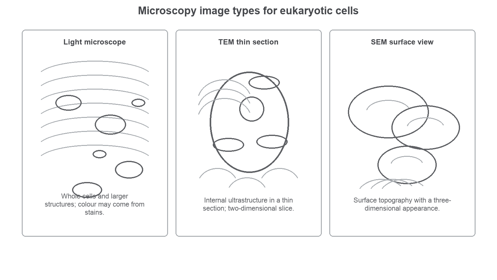
The diagram summarises the image evidence you should look for when deciding which microscope produced an image.
[DIAGRAM: microscopy_image_types: Lesson 012: Microscopy image types for eukaryotic cells - diagram 01; asset_slug: 012_m02_1_1_microscopy_to_observe_eukaryotic_cells__diagram_01; recommended_method: drawn_biology; description: A clean 16:9 three-panel schematic comparing light microscope, TEM and SEM image styles: whole cells/larger structures, a thin-section internal ultrastructure view, and a surface/topography view with three-dimensional appearance.]
| Microscope | Image style | Best evidence for |
|---|---|---|
| Light microscope | Whole cells, larger structures, colour if naturally pigmented or stained | Cell arrangement, cell shape, larger visible structures. |
| TEM | Thin, two-dimensional internal section | Internal ultrastructure and organelle detail. |
| SEM | Surface view with three-dimensional appearance | Cell or tissue surface shape and topography. |
Electron microscopy normally uses dead, fixed specimens in a vacuum, so it is not the right tool for observing a living cell in real time. It is also not enough to say "electron microscope" if the question asks for image type. TEM and SEM answer different questions.
Choosing The Microscope
In OCR-style questions, the mark usually comes from the reason, not just the microscope name. A good answer links the microscope to the evidence needed.
Matching method to evidence
A biologist wants to investigate the internal structure of a mitochondrion in an animal cell.
Choice: TEM.
Reasoning: the question is about internal ultrastructure, so the image needs a thin section through the cell or organelle. A TEM can produce a two-dimensional image showing internal detail. An SEM would mainly show the surface of the specimen, and a light microscope would not show the fine internal detail needed.
Use a short decision sequence:
- Identify the structure or feature being investigated.
- Decide whether the useful evidence is whole-cell layout, internal detail, or surface detail.
- Name the microscope that produces that kind of image.
- State one limitation so you do not overclaim.
| Investigation aim | Best choice | Good justification |
|---|---|---|
| Compare the shapes of cells in a thin plant tissue section | Light microscope | Shows whole cells and larger visible structures. |
| See internal membranes inside an organelle | TEM | Produces a thin-section image of internal ultrastructure. |
| Examine the surface of a pollen grain or epithelial cell | SEM | Shows surface topography with a three-dimensional appearance. |
This is also a How Science Works idea. Improved microscope images have changed cell models over time: biologists moved from seeing cells as simple compartments to building more detailed explanations of cell structure and organelles. The image is evidence, and the model changes when the evidence improves.
Interpreting Image Limits
Every microscope image has limits. Interpreting those limits is part of good Biology, because a micrograph is evidence produced by a method.
Do not overclaim from a light microscope image. If a small structure is not resolved, you cannot confidently label it just because you know from theory that it exists. You can say that a larger structure such as a nucleus or chloroplast is visible when the image supports that observation, but detailed organelle ultrastructure needs electron microscopy.
Do not overclaim from an electron micrograph either. Electron microscope images are usually greyscale, although published versions may be false-coloured. False colour can help the viewer distinguish structures, but it is not the natural colour of the specimen. TEM and SEM preparation can also alter or damage specimens, so the image should be interpreted as prepared evidence rather than a living cell exactly as it was.
For this OCR row, learn the image evidence: light microscope for whole cells and larger visible structures, TEM for internal thin-section detail, and SEM for surface/topography. Exact magnification and resolution values are not required here.