2.1.1(k) - Prokaryotic and eukaryotic cell comparison
This lesson is about comparing two basic kinds of cellular organisation. You are not learning every organelle again; you are learning the precise structural features that let a biologist distinguish a prokaryotic cell from a eukaryotic cell. The row also links to microscopy, so the lesson includes one scale calculation using the OCR magnification relationship.
Comparison Boundary
Biologists classify cells into prokaryotic cells and eukaryotic cells by how the cell is organised internally. The central idea is not that one cell type is "simple" and the other is "better". The central idea is where the genetic material is, whether the cell has membrane-bound compartments, and which structures are shared.
Prokaryotic cell
A cell that lacks a membrane-bound nucleus and lacks membrane-bound organelles; its DNA is found in the cytoplasm, usually in a region called the nucleoid.
Eukaryotic cell
A cell with DNA enclosed in a membrane-bound nucleus and with membrane-bound organelles such as mitochondria, endoplasmic reticulum and Golgi apparatus.
Structure means the arrangement of visible parts of the cell. Ultrastructure means finer cellular detail, usually the detail revealed by electron microscopy. In this lesson, the key ultrastructure idea is the contrast between a cell without internal membrane-bound compartments and a cell with them.
All living cells still share a core set of features:
| Feature shared by cells | Why it matters |
|---|---|
| Cell surface membrane | Separates the cell contents from the outside and controls exchange. |
| Cytoplasm | Contains enzymes and is where many metabolic reactions occur. |
| DNA | Carries genetic information. |
| Ribosomes | Are the site of protein synthesis. |
Core Structures
The diagram summarises the comparison using a typical bacterial prokaryotic cell and a typical eukaryotic animal cell. Treat it as a map of the comparison, not as a claim that every prokaryote or every eukaryote has every optional surface feature shown.
[DIAGRAM: prokaryotic_eukaryotic_comparison: Lesson 017: Prokaryotic and eukaryotic cell comparison - diagram 01; asset_slug: 017_m02_1_1_prokaryotic_and_eukaryotic_cell_comparison__diagram_01; recommended_method: drawn_biology; description: A clean side-by-side line diagram of a typical prokaryotic cell and a typical eukaryotic animal cell, with labels for shared features and OCR-safe differences including cell surface membrane, cytoplasm, DNA/nucleoid or nucleus, ribosome size, plasmid, cell wall and membrane-bound organelles.]
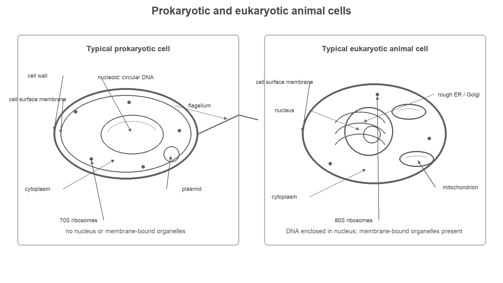
A comparison answer should be built from paired statements:
| Feature | Typical prokaryotic cell | Eukaryotic cell |
|---|---|---|
| Nucleus | No membrane-bound nucleus. | Has a membrane-bound nucleus. |
| DNA arrangement | DNA is usually circular and lies in the cytoplasm, often in a nucleoid region. | DNA is arranged as linear chromosomes inside the nucleus. |
| Plasmids | Small circular plasmids may be present. | Plasmids are not a normal feature of animal or plant cells at this level. |
| Membrane-bound organelles | Absent. | Present, for example mitochondria, ER and Golgi apparatus. |
| Ribosomes | Smaller ribosomes, commonly described as 70S. | Larger cytoplasmic ribosomes, commonly described as 80S. |
| Cell wall | Typical bacterial cells have a cell wall containing murein, also called peptidoglycan. | Plant and fungal cells may have cell walls, but animal cells do not. Plant walls contain cellulose. |
| Size | Usually smaller, often about 0.1-5 um. | Usually larger, often about 10-100 um. |
The phrase "typical bacterial cell" matters. Some bacterial cells have a capsule, pili or a flagellum, but those features are not universal. OCR comparison marks are more likely to reward the stable distinctions: nucleus, membrane-bound organelles, DNA form, ribosome size, cell wall material and relative size.
Compartmentation
The most important difference is compartmentation. A membrane-bound organelle is a cell structure surrounded by its own membrane. Eukaryotic cells use these organelles to separate different jobs into different spaces.
For example, mitochondria carry out aerobic respiration in eukaryotic cells, while the rough endoplasmic reticulum and Golgi apparatus are involved in making and processing proteins. You do not need the full sequence of protein secretion in this lesson, but you do need to recognise that those membrane-bound structures are eukaryotic features.
Prokaryotic cells do not have a nucleus, mitochondria, chloroplasts, ER or Golgi apparatus. Their DNA, ribosomes and many enzymes are in the cytoplasm. Because the cell is much smaller, substances can diffuse over shorter internal distances, so prokaryotic cells do not need the same internal compartment system to be OCR-identifiable as cells.
In a comparison question, do not write only "eukaryotic cells have organelles". Say "eukaryotic cells have membrane-bound organelles, whereas prokaryotic cells do not". Ribosomes are present in both cell types.
Walls, Ribosomes And Scale
Ribosomes are not surrounded by a membrane, so they are found in both cell types. The comparison is their size: prokaryotic ribosomes are smaller, commonly called 70S ribosomes, while eukaryotic cytoplasmic ribosomes are larger, commonly called 80S ribosomes. The "S" is a sedimentation unit, so it should not be treated like an ordinary length or mass unit.
Cell walls are another place where students often lose precision. A typical bacterial prokaryotic cell has a cell wall containing murein, also called peptidoglycan. Some eukaryotic cells have cell walls, but not animal cells. Plant cell walls contain cellulose, so "has a cell wall" on its own is not enough to distinguish a bacterium from every eukaryotic cell.
Scale can support identification in micrographs. Prokaryotic cells are usually much smaller than eukaryotic cells, though there is overlap in real biological examples. Use size as supporting evidence, not as the only proof.
Magnification
Use the same unit for image size and real object size before substituting. A common safe route is to convert millimetres to micrometres using:
Calculating magnification of a bacterial cell image
A bacterial cell has an actual length of 2.0 um. Its length in a photomicrograph is 18 mm.
Step 1: Convert the image size into micrometres.
Step 2: Substitute into the OCR magnification relationship.
The image is magnified x9000. This does not mean the real bacterium is 9000 um long; it means the image is 9000 times larger than the real object.
Exam Comparison
The command word Compare means you should give similarities and differences. For this lesson, a strong answer is usually a set of direct paired contrasts, not a long description of one cell followed by a long description of the other.
Good comparison language looks like this:
| Weak answer | Stronger answer |
|---|---|
| Prokaryotes have no nucleus. | Prokaryotic cells have no membrane-bound nucleus, whereas eukaryotic cells have DNA enclosed in a nucleus. |
| Eukaryotes have organelles. | Eukaryotic cells have membrane-bound organelles such as mitochondria, whereas prokaryotic cells lack membrane-bound organelles. |
| Prokaryotes are smaller. | Prokaryotic cells are usually smaller than eukaryotic cells, so a very small cell with no nucleus and no membrane-bound organelles is likely to be prokaryotic. |
| Bacteria have cell walls. | Typical bacterial cell walls contain murein/peptidoglycan; plant eukaryotic cell walls contain cellulose, and animal cells lack cell walls. |
When using a described micrograph, combine evidence. A small cell with no visible nucleus and no membrane-bound organelles suggests a prokaryotic cell. A larger cell with a nucleus and mitochondria suggests a eukaryotic cell. Size alone is not as strong as a structural feature, because measurements vary and images may show only part of a cell.
Quick Check
Use this as a short comprehension check on the section above.
One final check is to test the precision of the shortcut you use in your own head.