2.1.2(q) - Chemical tests for biological molecules
This lesson teaches how to carry out and interpret the four qualitative tests named in OCR Biology A for biological molecules. You will learn what each reagent shows, how to keep the method safe and valid, and how to turn a colour change into a careful biological conclusion without overclaiming.
Qualitative test logic
A qualitative test tells you whether a substance is detected, usually through a visible colour change or visible precipitate/emulsion. It does not, by itself, give an exact concentration. That is why a good answer separates the observation from the conclusion: "the solution turned purple" is the observation; "protein is present" is the conclusion.
Before testing foods or tissue extracts, the sample is usually ground or finely broken up, then mixed with water to make a solution or fine suspension. This increases contact between the biological molecules and the reagent. Starch is a small exception: iodine solution can be added directly to a solid specimen as well as to a suspension.
Two control tubes make the results more trustworthy:
| Control | What it contains | Why it matters |
|---|---|---|
| Positive control | A known sample containing the molecule being tested for | Checks the reagent and conditions can give the expected positive result |
| Negative control | Distilled water or a known sample without the molecule | Checks the reagent or apparatus has not contaminated the test |
A chemical test is only useful if the method, controls and observation all support the conclusion.
Protein and starch tests
The Biuret test detects protein because peptide bonds in proteins give a mauve, lilac, violet or purple colour in alkaline copper sulfate conditions. In school practical work, Biuret reagent may already contain the alkali and copper sulfate. A careful method is to add alkali to the protein solution first, then add a small amount of copper sulfate and mix gently. The positive colour may appear slowly, so do not dismiss a tube immediately after mixing.
The iodine test detects starch. Iodine solution in potassium iodide is brown or orange-brown before testing. A positive starch result is a blue-black colour. If the iodine remains pale brown or orange-brown, the result supports that no starch, or very little starch, was detected.
| Molecule tested | Reagent | Main method point | Positive observation |
|---|---|---|---|
| Protein | Biuret reagent or alkali plus copper sulfate | Mix gently; no heating needed | Mauve, lilac, violet or purple |
| Starch | Iodine solution in potassium iodide | Add to solution, suspension or solid specimen | Blue-black |
When a question asks you to "interpret" a test result, name both the observation and the molecule detected. "Purple" alone is not the conclusion; "purple, so protein is present" is.
Reducing and non-reducing sugar tests
Benedict's test detects reducing sugars such as glucose. Add Benedict's reagent to the sample, mix, then heat the tube in a boiling water bath for about five minutes. Use a water bath rather than a direct flame because the mixture can spit or bump violently if heated directly.
A negative Benedict's result remains blue. A positive result forms an insoluble precipitate, with the colour depending roughly on how much reducing sugar is present: green, yellow, orange, then brick red or red-brown as the amount increases.
Non-reducing sugars need an extra sequence. They may give a negative direct Benedict's test because they do not reduce Benedict's reagent before hydrolysis. To test for them, first carry out the direct Benedict's test. If it is negative, heat a fresh sample with dilute hydrochloric acid to hydrolyse the sugar, neutralise the solution with sodium hydrogencarbonate or another suitable alkali, check with pH paper, then repeat Benedict's test.
Interpreting a non-reducing sugar test
A food extract remains blue after the direct Benedict's test. A fresh sample is then hydrolysed with dilute acid, neutralised, and retested with Benedict's reagent. This time an orange precipitate forms.
The first result shows no reducing sugar was detected in the original extract. The positive result after hydrolysis shows reducing sugars were produced by breaking down another sugar. Together, those results support the presence of a non-reducing sugar in the original extract.
Neutralisation is not a decorative step. Benedict's test is carried out in alkaline conditions, so acid left after hydrolysis can interfere with the repeated test. The OCR-safe logic is: direct test first, hydrolyse if needed, neutralise, then retest.
Lipid emulsion test
The emulsion test detects lipids. Shake the sample with ethanol for about a minute so that any lipid dissolves in the ethanol. Then add the ethanol extract to water, leaving the solid material behind where possible. If lipid is present, a white cloudy or milky emulsion forms.
This result is an emulsion or suspension, not a precipitate. That language matters because OCR mark schemes often distinguish the lipid emulsion from the precipitate in Benedict's test.
Ethanol is flammable, so keep it away from naked flames and use a bung when shaking the tube. If a sample contains both water and ethanol, the order matters: lipid dissolves in ethanol first, then becomes dispersed when the ethanol is added to water.
PAG9 interpretation
In this OCR practical skill, the useful habit is to record the test condition, observation and conclusion separately. That makes it easier to spot when a method is incomplete or when a student has overinterpreted a colour.
[DIAGRAM: biological_molecule_test_results_map: Lesson 27: Chemical tests for biological molecules - diagram 01; asset_slug: 027_m02_1_2_chemical_tests_for_biological_molecules__diagram_01; recommended_method: drawn_biology; description: A clean 16:9 grey method-to-conclusion map linking Biuret, Benedict's direct and hydrolysis retest, iodine, and ethanol emulsion tests to their positive observations and biological conclusions.]
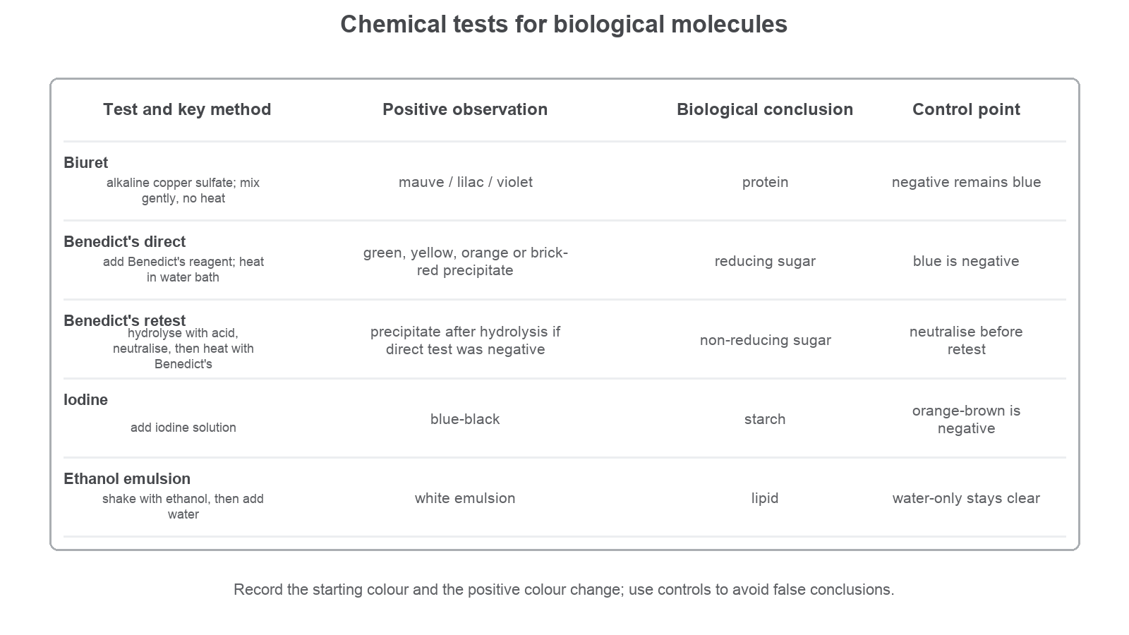
The diagram is a summary map, not a substitute for the method. Use it after you can already explain why each result leads to its conclusion.
Unknown sample results
A blended seed extract gives these results:
| Test | Observation |
|---|---|
| Biuret | Purple after gentle mixing |
| Benedict's direct test | Blue after heating |
| Benedict's after acid hydrolysis and neutralisation | Orange precipitate after heating |
| Iodine | Brown |
| Emulsion | White cloudy emulsion |
The sample contains protein because Biuret turned purple. It contains a non-reducing sugar because the direct Benedict's test was negative but the hydrolysed, neutralised retest was positive. It does not have starch detected by iodine because iodine stayed brown. It contains lipid because a white cloudy emulsion formed.
The listed tests do not detect every biological molecule. For example, cellulose, vitamins and inorganic ions are not identified by this set of four tests. A negative result only means that the specific molecule was not detected under the test conditions.