4.2.2.2 - The Heart and Blood Vessels
The circulatory system moves substances around the body quickly, so cells can receive oxygen and glucose and remove carbon dioxide. In humans, the heart is the muscular organ that pumps blood through blood vessels. This lesson focuses on the AQA boundary: the double circulatory system, the named vessels connected to the heart, lung gas exchange, pacemakers, the three vessel types and simple blood-flow rate calculations.
Double circulation
The heart is an organ: it is made of tissues working together to pump blood. Humans have a double circulatory system, which means blood passes through the heart twice during one complete circuit of the body.
Double circulatory system
A circulation system in which blood travels from the heart to the lungs and back to the heart, then from the heart to the rest of the body and back again.
The right ventricle pumps blood to the lungs. In the lungs, gas exchange takes place: oxygen enters the blood and carbon dioxide leaves the blood. The blood then returns to the heart before the left ventricle pumps it around the rest of the body.
This is why the two ventricles have different jobs:
| Heart chamber | Main pumping job |
|---|---|
| Right ventricle | Pumps blood to the lungs |
| Left ventricle | Pumps blood around the rest of the body |
The left ventricle pumps to a much longer circuit than the right ventricle, so it must generate enough pressure to send blood around the body. Do not describe this as the heart "making" oxygen. Oxygen is added to the blood at the gas-exchange surface in the lungs.
Named vessels
AQA limits the named blood vessels associated with the heart to the aorta, vena cava, pulmonary artery, pulmonary vein and coronary arteries.
| Vessel | What it does in this lesson |
|---|---|
| Vena cava | Carries blood from the body back to the heart |
| Pulmonary artery | Carries blood from the right ventricle to the lungs |
| Pulmonary vein | Carries blood from the lungs back to the heart |
| Aorta | Carries blood from the left ventricle to the rest of the body |
| Coronary arteries | Supply the heart muscle with blood |
[DIAGRAM: asset_name: double-circulation-and-named-vessels - diagram 1; asset_slug: 021_4_2_2_2_the_heart_and_blood_vessels_diagram1; file: diagram_assets/imagegen_regen_all/021_4_2_2_2_the_heart_and_blood_vessels_diagram1_imagegen.png; recommended_method: codex_image_gen; description: Monochrome flow diagram of the double circulatory system showing right atrium, right ventricle, left atrium, left ventricle, vena cava, pulmonary artery, lungs with trachea, bronchi, alveoli and capillary network, pulmonary vein, aorta, body tissues and coronary arteries supplying the heart muscle.]
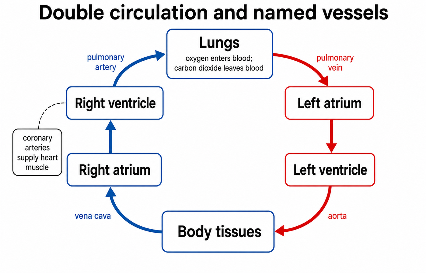
A common mistake is to define arteries and veins by oxygen level. That works for many vessels, but it fails for the pulmonary vessels. The safer GCSE rule is direction: arteries carry blood away from the heart, while veins carry blood towards the heart. The pulmonary artery carries blood away from the heart to the lungs, even though that blood has not yet picked up oxygen from the lungs.
Use vessel direction first: artery means away from the heart, and vein means towards the heart.
That direction rule is the easiest way to keep all four main routes straight.
Lungs and gas exchange
In this specification point, lung knowledge is restricted to the trachea, bronchi, alveoli and the capillary network around the alveoli.
Air travels down the trachea. The trachea branches into bronchi, which lead deeper into the lungs. At the ends of the airways are many tiny air sacs called alveoli. Gas exchange happens across the surface of the alveoli and the surrounding capillaries.
Alveoli
Tiny air sacs in the lungs where oxygen diffuses into the blood and carbon dioxide diffuses out of the blood.
Alveoli are adapted for gaseous exchange because there are many of them, giving a large surface area. Their walls are thin, and they are surrounded by a capillary network, giving a short diffusion distance between air and blood. Blood flow through the capillary network helps maintain a concentration gradient, because oxygenated blood is carried away and blood with less oxygen is brought to the exchange surface.
Pacemakers and heart rate
Heart rate is how many times the heart beats per minute. The natural resting heart rate is controlled by a group of cells in the right atrium that act as a pacemaker. These cells produce electrical impulses that help set the rhythm of the heartbeat.
Pacemaker
A group of cells in the right atrium that controls the natural resting heart rate.
If the heart rate is irregular, an artificial pacemaker can be used. Artificial pacemakers are electrical devices that correct irregularities in heart rate. At GCSE level, you do not need the detailed names of the heart's electrical pathway.
Vessel structure and function
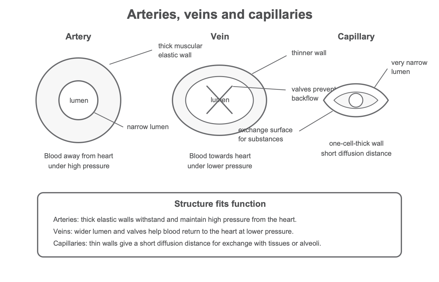
The body contains three different types of blood vessel: arteries, veins and capillaries. Their structures fit their functions.
[DIAGRAM: asset_name: artery-vein-capillary-structure - diagram 2; asset_slug: 021_4_2_2_2_the_heart_and_blood_vessels_diagram2; file: diagram_assets/imagegen_regen_all/021_4_2_2_2_the_heart_and_blood_vessels_diagram2_imagegen.png; recommended_method: codex_image_gen; description: Monochrome cross-section comparison of an artery with a thick muscular elastic wall and narrow lumen, a vein with thinner wall, wider lumen and valves, and a capillary with a one-cell-thick wall and very narrow lumen, with concise labels linking structure to function.]
Lumen
The space inside a blood vessel that blood flows through.
| Vessel type | Structure | How the structure relates to function |
|---|---|---|
| Artery | Thick muscular and elastic wall; relatively narrow lumen | Carries blood away from the heart under high pressure; the wall withstands and maintains pressure |
| Vein | Thinner wall; wider lumen; valves | Carries blood back to the heart under lower pressure; valves help prevent backflow |
| Capillary | Wall one cell thick; very narrow lumen; many branches | Allows exchange of substances with tissues or alveoli over a short diffusion distance |
When an exam question asks you to explain structure and function, link the feature to the job. "Arteries have thick walls" is only a description. "Arteries have thick muscular and elastic walls to withstand the high pressure of blood pumped from the heart" is an explanation.
Now practise turning those structure-function links into direct comparison language.
Blood flow rate
AQA can ask you to use simple compound measures such as rate. A blood-flow rate tells you the volume of blood moving through a vessel each unit of time.
Blood-flow rate
The unit depends on the volume unit and the time unit. For example, if volume is measured in cm^3 and time in minutes, the rate is measured in cm^3/min.
Worked example:
A blood vessel carries 480 cm^3 of blood in 6 minutes. Calculate the rate of blood flow.
- Write the relationship:
rate = volume ÷ time - Substitute the values:
rate = 480 cm^3 ÷ 6 min - Calculate:
rate = 80 cm^3/min
If the time is given in seconds, either keep the answer per second or convert the time before you calculate, depending on the unit asked for. The important step is to keep the volume unit and time unit together in the final answer.
Exam precision
For this lesson, keep your answers inside the AQA boundary. You do not need the names of the heart valves, although valves can be described as preventing backflow if their function is relevant. You do not need to explain coronary heart disease treatments, blood components, ECG traces or named parts of the heart's electrical pathway.
The most common precision trap is artery versus vein. Do not write that all arteries carry oxygenated blood or that all veins carry deoxygenated blood. The pulmonary artery and pulmonary vein are the exceptions, so direction relative to the heart is the safer definition.
A strong 4-mark explanation often follows this pattern:
The heart is a pump in a double circulatory system. The right ventricle pumps blood to the lungs, where gas exchange takes place at the alveoli and capillary network. The pulmonary vein returns blood to the heart. The left ventricle then pumps blood through the aorta to the rest of the body.