4.1.1.6b - Aseptic Technique and Zones of Inhibition
In this Biology-only required practical, you prepare an uncontaminated bacterial culture and test how well antiseptics or antibiotics inhibit bacterial growth. The result is judged from clear areas on an agar plate called zones of inhibition. The practical is really about two linked skills: keeping the culture uncontaminated, then collecting valid measurement data from the clear zones.
Why uncontaminated cultures matter
A culture is a population of microorganisms grown in a nutrient medium. Bacteria can be grown in a nutrient broth solution, or as visible colonies on nutrient agar in a Petri dish.
For this practical, the culture must be uncontaminated. That means the microorganisms growing on the agar should be the bacteria deliberately added, not microorganisms from the air, bench, hands, equipment or the outside of a container.
Aseptic technique
A set of methods used to prevent unwanted microorganisms entering or leaving a culture.
Contamination matters for two reasons. It can spoil the results because extra microorganisms may grow on the plate. It can also create a health risk if microorganisms from the culture escape into the surroundings or if harmful contaminants grow.
A valid inhibition-zone investigation depends on knowing that any bacterial growth came from the intended culture and any clear zone came from the antiseptic or antibiotic being tested.
Preparing an uncontaminated culture
The core aseptic steps all reduce contamination risk:
- Sterilise Petri dishes and culture media before use so there are no living microorganisms already present.
- Sterilise an inoculating loop by passing it through a flame before transferring microorganisms, so previous microorganisms on the loop are killed.
- Open lids for the shortest possible time and hold them at an angle, so fewer airborne microorganisms can fall onto the agar.
- Secure the Petri dish lid with adhesive tape, but do not seal it all the way around.
- Incubate school or college bacterial cultures at, or no higher than, 25 degrees C.
- Store agar plates upside down during incubation.
The tape reduces the chance that the lid will come off, so microorganisms are less likely to escape and the student is less likely to touch the culture. The lid is not sealed all the way around because complete sealing can create anaerobic conditions inside the plate.
Plates are incubated upside down so condensation does not drip onto the agar and spread bacteria across the surface. The 25 degrees C school temperature reduces the chance that harmful human pathogens will grow well; 37 degrees C is close to human body temperature and is not used for this school practical.
Testing antiseptics or antibiotics
The practical tests the effect of antiseptics or antibiotics on bacterial growth. A simple hypothesis might be: "If an antiseptic is more effective, then it will produce a larger clear zone on the bacterial lawn."
A typical method is:
- Disinfect the bench, wash hands, and set up a Bunsen burner on a heatproof mat.
- Label the underside of a sterile agar plate, not the lid, so labels stay aligned with the agar.
- Add a small volume of bacterial culture to the agar plate using aseptic technique.
- Spread the bacteria across the agar to make a bacterial lawn.
- Place sterile filter paper discs soaked in different antiseptics on marked positions, or use commercially prepared antibiotic discs.
- Secure the lid with small pieces of tape, keeping it closed but not fully sealed.
- Incubate the plate upside down at 25 degrees C for a set time, often about 48 hours.
- Without opening the lid, measure the clear zone around each disc.
[DIAGRAM: asset_name: plate_zone_measurement: Agar plate layout and zone measurements - diagram 1; asset_slug: 008_4_1_1_6b_required_practical_2_aseptic_technique_and_inhibition_zones_diagram1; file: diagram_assets/008_4_1_1_6b_required_practical_2_aseptic_technique_and_inhibition_zones_diagram1.png; recommended_method: deterministic_drawn; description: Top-down monochrome nutrient agar plate divided into sectors with three antimicrobial discs, clear zones, measurement diameters at 90 degrees, and an area calculation callout.]
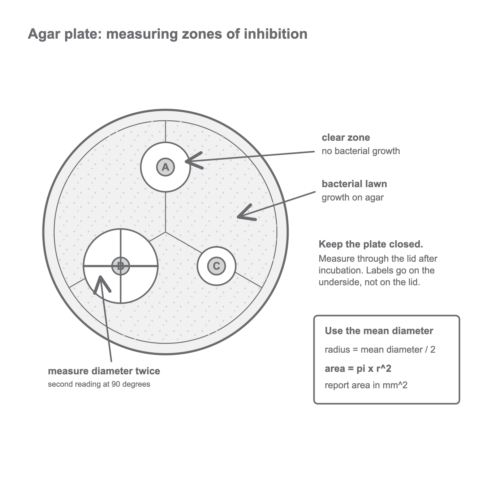
The independent variable is the antiseptic or antibiotic used, or the concentration if one substance is tested at different concentrations. The dependent variable is bacterial growth, measured using the diameter or area of the zone of inhibition. Control variables include the species of bacteria, agar type, incubation temperature, incubation time, disc size and volume or concentration of antimicrobial substance on each disc.
Measuring zones and calculating area
A zone of inhibition is the clear area where bacteria have not grown around an antiseptic or antibiotic disc. A larger clear zone usually means stronger inhibition under the same test conditions.
Be careful with the word "usually". Zone size is good evidence only when the comparison is fair. Different substances may diffuse through agar at different rates, so you should compare results from the same method, same bacterial lawn and same incubation conditions.
Clear zones are not always perfect circles. Measure the diameter twice, with the second measurement at 90 degrees to the first. Then calculate the mean diameter.
Area of a circular zone
Here, is the radius of the zone. If the mean diameter is in mm, divide by 2 to find the radius in mm. The area will be in mm^2.
Worked example:
A clear zone has diameter readings of 20 mm and 18 mm.
Mean diameter:
Radius:
Area:
To three significant figures, the area is 283 mm^2.
Judging quality of evidence
A good conclusion uses the data and keeps the method limitations in mind. For example, "Antiseptic B was most effective in this test because it produced the largest mean clear-zone area" is stronger than "Antiseptic B is always the best".
Reliability improves if each antiseptic or antibiotic is tested more than once and a mean is calculated. If one result is very different from the others, check whether it is anomalous before including it in a mean.
Validity improves when the method really tests the intended variable. A control disc with sterile water can show whether the paper disc or water alone causes a clear zone. Keeping the lid closed while measuring reduces the risk of releasing bacteria and keeps the practical safe.
Risk reduction is part of the practical skill, not an optional extra. Clean benches and hands before and after the experiment, use aseptic technique throughout, keep plates closed after incubation, handle antibiotic discs with forceps, and sterilise or safely dispose of anything that touched the microorganisms.
The same logic is used when judging whether the measurements are reliable enough to support a conclusion.