4.1.1.5 - Microscopy, Magnification and Resolution
Microscopes let biologists study cells and the structures inside them. As microscope technology improved, biologists could make images bigger and, more importantly, see finer detail. This lesson focuses on how light and electron microscopes differ, and how to handle magnification calculations without losing the units.
Why microscopes developed
Early microscopes let scientists see that living organisms are made of cells. Over time, microscopy techniques improved, so cells could be studied at higher magnification and with better resolution.
The important GCSE idea is that better technology can give scientists better evidence. When biologists could see more detail inside cells, they could build more accurate explanations of sub-cellular structures.
This does not mean every microscope works in the same way. A light microscope is useful for viewing cells, but an electron microscope can show much finer detail. The specification limits the comparison to two features: magnification and resolution.
Sub-cellular structure
A sub-cellular structure is a structure inside a cell, such as a nucleus, mitochondrion or ribosome.
In this lesson, the point is not to learn a new list of cell parts. The point is that better microscopes made smaller cell details visible enough to study.
Magnification and resolution
Magnification and resolution are not the same thing.
Magnification
Magnification is how many times larger the image is than the real object.
If a cell is shown at x400, the image is 400 times larger than the real cell. Magnification makes the image bigger, but it does not guarantee that the image is clear.
Resolution
Resolution is the ability to distinguish two points that are close together as separate points.
High resolution gives a clearer image with more detail. If resolution is poor, two close structures may blur into one shape even if the image is enlarged. This is why increasing magnification alone can produce a bigger blurry image rather than a useful one.
[DIAGRAM: asset_name: magnification_resolution_comparison - diagram 1; asset_slug: 006_4_1_1_5_microscopy_magnification_and_resolution_diagram1; file: diagram_assets/imagegen_regen_all/006_4_1_1_5_microscopy_magnification_and_resolution_diagram1_imagegen.png; recommended_method: codex_image_gen; description: Monochrome deterministic comparison of a light microscope-style view and an electron microscope-style view of the same simple cell area. Show the light microscope panel with lower magnification and lower resolution, where two close sub-cellular dots merge into a blurred shape; show the electron microscope panel with higher magnification and higher resolving power, where the close dots are separate. Include labels for magnification, resolution and resolving power. Deterministic drawing is required because the assessed distinction depends on labels and geometry.]
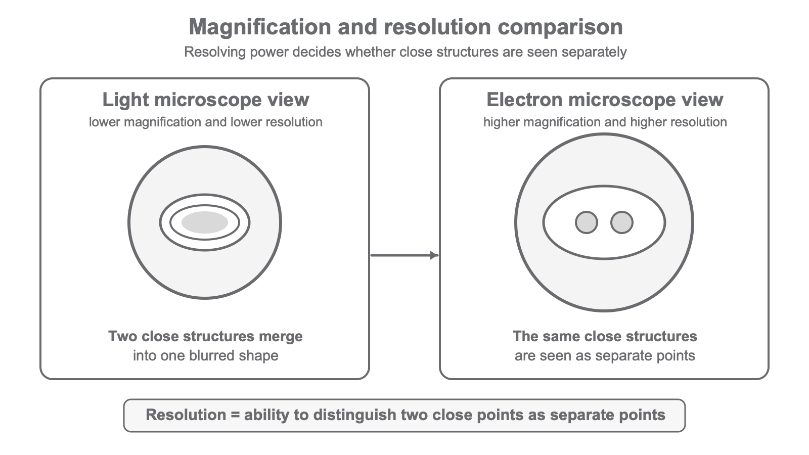
Light and electron microscopes
A light microscope uses light to form an image. Light microscopes are useful for viewing whole cells and some larger cell structures, but their magnification and resolving power are limited.
An electron microscope has much higher magnification and much higher resolving power than a light microscope. This means it can be used to study cells in much finer detail.
Electron microscopy has increased understanding of sub-cellular structures because it lets biologists see smaller structures more clearly. For example, structures such as ribosomes can be studied in much more detail using electron microscopy than using a light microscope.
The comparison you need is precise:
- electron microscopes have higher magnification than light microscopes
- electron microscopes have higher resolution, or higher resolving power, than light microscopes
- higher resolving power means finer detail can be seen
- seeing finer detail has helped biologists understand more sub-cellular structures
A comparison answer should use direct comparative wording, such as higher than, lower than or more detail than.
The magnification equation
Magnification calculations use three quantities:
magnificationsize of imagesize of real object, also called real size or actual size
Magnification
Magnification has no unit because it is a ratio. The image size and real object size must be in the same unit before you divide them.
Worked example:
A cell has a real diameter of 0.05 mm. Its image has a diameter of 20 mm.
- Write the relationship:
magnification = size of image / size of real object - Substitute:
magnification = 20 / 0.05 - Calculate:
magnification = 400 - Give the answer:
x400
To find real size, rearrange the equation:
size of real object = size of image / magnification
Worked example:
A micrograph image of a cell is 18 mm wide. The magnification is x600. Calculate the real width of the cell in micrometres.
- Rearrange:
real size = image size / magnification - Substitute:
real size = 18 mm / 600 - Calculate:
real size = 0.03 mm - Convert:
0.03 mm x 1000 = 30 um - Answer:
30 um
Units, standard form and checks
Microscopy calculations often use very small lengths, so unit conversion matters. At GCSE, the useful prefixes here are:
centi, symbolc, means10^-2milli, symbolm, means10^-3micro, often shown asuin plain text, means10^-6nano, symboln, means10^-9
For length units:
1 cm = 10 mm1 mm = 1000 um1 um = 1000 nm
You may also need standard form. Standard form writes a number as a value from 1 up to but not including 10, multiplied by a power of 10.
Examples:
3000 nm = 3.0 x 10^3 nm0.000002 m = 2.0 x 10^-6 m25 um = 2.5 x 10^1 um
[DIAGRAM: asset_name: magnification_equation_units - diagram 2; asset_slug: 006_4_1_1_5_microscopy_magnification_and_resolution_diagram2; file: diagram_assets/006_4_1_1_5_microscopy_magnification_and_resolution_diagram2.png; recommended_method: deterministic_drawn; description: Monochrome deterministic formula-and-units visual showing magnification = size of image / size of real object, rearranged forms for image size and real size, and a unit conversion ladder from cm to mm to um to nm with x10, x1000 and x1000 steps. Include a small reminder that magnification has no unit and image size and real size must be in the same unit before division. Deterministic drawing is required because the formulas and conversions are assessed.]
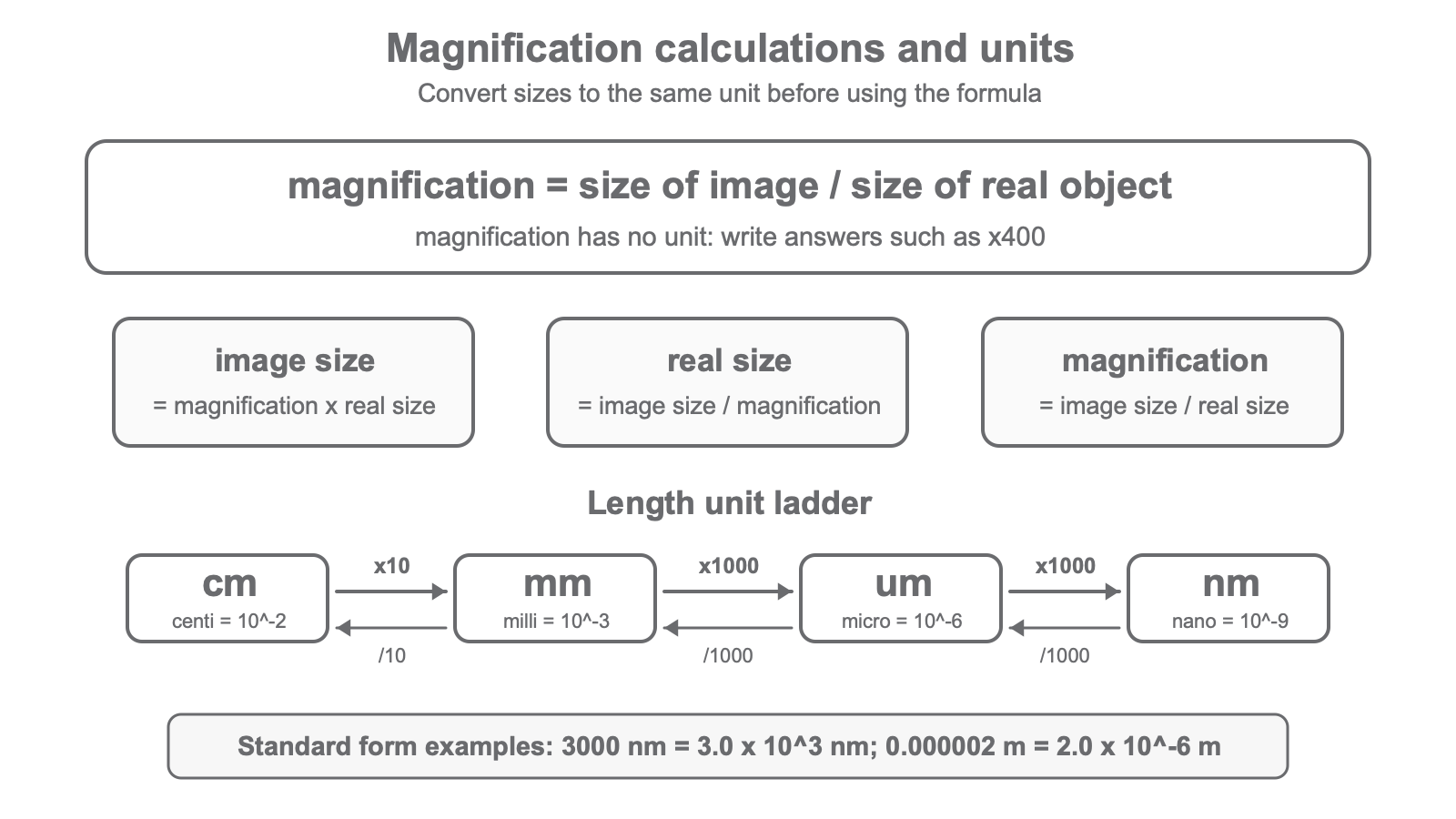
Use this exam routine:
- Identify which quantity the question asks for.
- Write the formula or rearranged formula.
- Convert the two size measurements into the same unit if needed.
- Substitute the numbers.
- Calculate carefully.
- Add a unit for real size or image size. Do not add a unit to magnification.
- Use standard form if the answer is very large or very small, or if the question asks for it.
Most magnification mistakes come from mixing units or treating magnification as if it had a unit.
The safest habit is to convert first, then calculate.