2.2.6 Brain Plasticity and Functional Recovery
The brain was once thought to be a fixed organ — fully developed by early childhood and incapable of meaningful change thereafter. We now know this is wrong. The brain continues to modify its structure and function throughout life in response to experience, learning, and injury. This capacity is called plasticity. A particularly dramatic example of plasticity occurs after brain trauma, when undamaged regions may take over the roles of destroyed tissue — a process known as functional recovery. This lesson examines the evidence for both phenomena, the mechanisms that underpin functional recovery, and the strengths and limitations of the research in this area.
Part 1 — Brain Plasticity
Plasticity
The brain's ability to change and adapt its structure and function throughout life in response to experience, learning, and trauma. This typically involves the formation of new synaptic connections and the strengthening or pruning of existing ones.
During infancy, the brain undergoes an explosion of synaptic growth, peaking at approximately 15,000 connections per neuron by 2–3 years of age — roughly double the number found in the adult brain. As we develop, connections that are frequently used become stronger while those that are rarely activated are eliminated. This process is known as synaptic pruning.
Synaptic Pruning
The process by which excess neurons and synaptic connections are eliminated in order to increase the efficiency of neuronal transmission. Frequently used pathways are strengthened while infrequently used pathways are removed.
For many years, researchers believed that a "critical window" for neural development existed in the first few years of life, after which the brain's structure was essentially fixed. Modern neuroscience has overturned this view. We now understand that synaptic pruning enables lifelong plasticity — new neural connections continue to form whenever the brain faces new demands, whether through learning a new skill, acquiring knowledge, or recovering from injury.
Research into plasticity
The most well-known demonstration of adult brain plasticity comes from the work of Eleanor Maguire and colleagues.
Maguire et al. (2000) — Used MRI scans to compare the brains of London taxi drivers with those of matched controls. Taxi drivers must pass an extremely demanding test called "The Knowledge," which requires memorising thousands of streets and routes across London. The researchers found that taxi drivers had significantly greater grey matter volume in the posterior hippocampus — a region associated with spatial navigation and memory — compared to controls. Furthermore, the volume of the posterior hippocampus showed a positive correlation with the number of years spent driving a taxi: the longer a driver had been in the job, the larger this brain region was. This was a correlational study using structural MRI, and Maguire concluded that the extensive spatial learning required by taxi driving physically altered the structure of the hippocampus.
Further evidence for experience-driven plasticity was provided by Draganski et al. (2006), who used brain imaging to scan medical students three months before and three months after their final examinations. Learning-induced structural changes were observed in the posterior hippocampus and the parietal cortex, presumably as a result of the intensive revision period. This demonstrates that even relatively short periods of focused learning can produce measurable changes in brain structure.
Priya has been learning to play the violin since the age of seven and practises for two hours every day. A neuroscientist scanning her brain notices that the area of her motor cortex corresponding to her left hand — the hand she uses to finger the strings — is significantly larger than the same region in non-musicians of her age. This illustrates brain plasticity: Priya's years of repetitive, skilled practice have caused structural changes in the regions of her brain responsible for fine motor control.
Part 2 — Functional Recovery after Brain Trauma
Functional Recovery
A form of neural plasticity in which, following damage through trauma (such as a stroke or head injury), the brain redistributes or transfers functions previously carried out by the damaged area(s) to other undamaged area(s), enabling some degree of continued functioning.
Functional recovery represents one of the most remarkable demonstrations of plasticity. When areas of the brain are damaged — through stroke, traumatic brain injury, surgery, or disease — healthy regions can sometimes compensate for the lost functions. Neuroscientists suggest that this process can occur rapidly in the initial period after trauma (spontaneous recovery) and then slows down over the following weeks and months. After this point, further improvement typically requires rehabilitative therapy.
Mechanisms of functional recovery
Several structural and physiological changes in the brain support functional recovery. The diagram below summarises four major mechanisms, and a fifth important process — unmasking of secondary neural pathways — is introduced immediately afterwards:
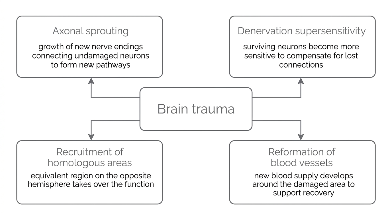
- Axonal sprouting — the growth of new nerve endings which connect with undamaged nerve cells, forming new neuronal pathways around the site of injury. This is analogous to finding an alternative route when a road is blocked.
- Denervation supersensitivity — when axons performing a particular function are destroyed, the surviving neurons carrying out similar work become more highly sensitive to compensate. However, this can sometimes produce negative effects such as heightened pain sensitivity.
- Recruitment of homologous areas — the equivalent region on the opposite hemisphere of the brain takes over the function of the damaged area. For example, if Broca's area (responsible for speech production) is damaged in the left hemisphere, the corresponding region in the right hemisphere may assume its function.
- Reformation of blood vessels — new blood supply develops around the damaged area as part of the brain's haemodynamic response, supporting the metabolic needs of recovering neurons.
- Unmasking of secondary neural pathways — neural circuits that normally lie dormant are activated to take over functions that can no longer be carried out by the damaged area. This is sometimes described through the law of equipotentiality, whereby secondary circuits surrounding the damaged region become active.
These mechanisms mean that function is not always permanently lateralised to a specific hemisphere — the brain has a degree of flexibility in where it processes information.
Case study evidence for functional recovery
One striking example of functional recovery involves a young girl named Jodi Miller, who underwent a hemispherectomy — the surgical removal of her entire right hemisphere — to control severe epileptic seizures. Despite losing half of her brain, Jodi was still able to control the left side of her body through the remaining left hemisphere. Her case demonstrates the power of axonal sprouting, recruitment of homologous areas, and reformation of blood vessels in enabling the brain to compensate for massive structural loss. This is an example of positive plasticity, where the brain's adaptive changes produce beneficial outcomes.
Part 3 — Negative Plasticity
Not all plasticity is beneficial. The brain's capacity for reorganisation can sometimes produce harmful or distressing outcomes — a phenomenon known as negative plasticity.
Phantom limb syndrome
Ramachandran and Hirstein (1998) — Investigated phantom limb syndrome, a condition experienced by 60–80% of amputees in which they continue to feel sensations — often painful — in a limb that has been removed. Ramachandran proposed that phantom limb pain is caused by cortical reorganisation in the somatosensory cortex: after amputation of a hand, sensory input from the face (which occupies an adjacent region of the somatosensory cortex) "invades" the deafferented hand zone. This means that touching the face can produce sensations that the brain interprets as coming from the missing hand. Ramachandran concluded that there is "tremendous latent plasticity even in the adult brain," but in this case the plasticity produces painful and distressing consequences rather than recovery.
Drug use and plasticity
Research has also demonstrated that prolonged drug use can alter brain structure in harmful ways. Medina et al. (2007) found evidence that the brain's adaptation to chronic substance use is associated with poorer cognitive functioning in later life and an increased risk of dementia. This represents another form of negative plasticity, where experience-driven neural changes produce lasting damage rather than improvement.
These findings highlight an important point: plasticity is a neutral mechanism. The brain adapts to whatever demands are placed upon it — whether those demands come from learning a new language or from chronic drug exposure. The outcome depends entirely on the nature of the experience.
After losing his left arm in an accident, David reports feeling intense tingling and pain in his missing hand whenever someone touches his face. His neurologist explains that this is phantom limb syndrome, caused by cortical reorganisation in the somatosensory cortex. The brain region that previously received input from David's left hand has been "invaded" by input from the adjacent face region. David's experience is an example of negative plasticity — his brain has adapted to the loss of sensory input, but the adaptation has produced painful rather than beneficial consequences.
The distinction between positive and negative plasticity is important for understanding that the brain's capacity for change is not inherently good or bad — it is a mechanism that responds to whatever input it receives. Recognising this helps explain why some forms of experience (such as chronic drug use or sensory deprivation) can lead to maladaptive neural changes.
Part 4 — Factors Affecting Functional Recovery
Cognitive reserve
Not everyone recovers equally from brain trauma. One factor that appears to influence the rate and extent of functional recovery is cognitive reserve — broadly defined as the level and duration of education a person has received.
Schneider et al. (2014) — Studied 8,769 patients with moderate to severe traumatic brain injury and tracked their recovery over one year. Of these, 214 patients achieved disability-free recovery (DFR). The researchers found that educational background was a significant predictor of recovery: 40% of those who achieved DFR had more than 16 years of education, compared to approximately 10% of those with fewer than 12 years. Schneider et al. concluded that higher cognitive reserve — built through extended engagement with education — may increase the brain's capacity for functional recovery, perhaps because educated individuals have developed more extensive neural networks that can compensate when some pathways are damaged.
The role of age
Age is another important factor. In general, younger brains show greater plasticity than older brains, and children tend to make more complete recoveries from brain injury than adults. However, research suggests that plasticity is not confined to childhood. Bezzola et al. (2012) demonstrated that 40 hours of golf training produced measurable changes in the neural representations of movement in participants aged 40–60. Using fMRI, the researchers observed increased motor cortex activity in the novice golfers compared to a control group, suggesting that experience-driven structural change continues well into middle age.
Limits of spontaneous recovery
It is important to recognise that functional recovery has limits. The brain can only "repair" itself to a certain extent through spontaneous mechanisms. Beyond this point, external intervention is typically needed. Liepert et al. (2000) demonstrated that constraint-induced movement therapy — in which stroke patients repeatedly practise using the affected limb while the unaffected limb is restrained — significantly improved motor performance. This finding has had important real-world applications in neurorehabilitation, showing that targeted therapy can extend recovery beyond what the brain achieves spontaneously.
Brain plasticity allows the brain to adapt throughout life, but functional recovery after trauma depends on multiple factors including age, cognitive reserve, and access to rehabilitative therapy. Spontaneous recovery has limits, and external intervention is often needed to maximise outcomes.
Understanding these factors is clinically important: it helps medical professionals predict which patients are most likely to recover and when therapeutic intervention should begin. The interplay between spontaneous recovery and rehabilitation is a key theme in modern neurorehabilitation.
Part 5 — Supporting Research
Animal research into plasticity
Hubel and Wiesel (1970) — Conducted a laboratory experiment in which they sutured shut one eye of newborn kittens for a period of several months, periodically opening the eye and monitoring brain activity in the visual cortex. The researchers found that although the deprived eye received no visual input, activity was still detected in the corresponding visual cortex. However, the cortical neurons that would normally respond to the closed eye had been taken over by input from the open eye — a process now understood as the development of ocular dominance columns. Hubel and Wiesel noted that during a critical period of high susceptibility (around weeks four and five), even 3–4 days of eye closure led to a significant decline in the number of neurons that could be driven by both eyes. This study provides evidence that brain areas receiving no input can be colonised by adjacent, highly stimulated areas — demonstrating plasticity at the neural level.
This research raises important ethical issues. The kittens suffered permanent visual impairment as a result of the procedure, and modern ethical guidelines would make it difficult to replicate this study. However, the findings have been influential in understanding sensitive periods in brain development and have contributed to the treatment of conditions such as amblyopia (lazy eye) in children.
Stem cell research
Emerging research has explored whether stem cells could be used to enhance functional recovery. Banerjee et al. (2014) treated patients who had suffered a total anterior circulation stroke with stem cell therapy. All participants in this small trial showed recovery, compared to a typical recovery rate of just 4% for this type of stroke. However, the study used only five participants and had no control group, meaning firm conclusions cannot be drawn. This illustrates a common limitation of research into functional recovery: sample sizes tend to be very small because the specific conditions being studied are relatively rare and ethical constraints limit experimental manipulation.
Evaluation Bank (AO3)
Strength: Research into brain plasticity has strong real-world application in the field of neurorehabilitation. Understanding the specific mechanisms involved in functional recovery — such as axonal sprouting and recruitment of homologous areas — has directly informed therapeutic interventions. For example, Liepert et al. (2000) demonstrated that constraint-induced movement therapy significantly improved motor performance in stroke patients by exploiting the brain's capacity for plasticity. This shows that plasticity research is not merely theoretical but has practical value in improving patient outcomes after brain trauma. The translation from laboratory findings to clinical practice gives this area of research high external validity, and it illustrates how psychological knowledge can be applied to benefit individuals (a positive ethical implication of research).
Limitation: Much of the evidence for functional recovery relies on case studies of individuals with rare or unusual brain injuries, such as Jodi Miller's hemispherectomy. While case studies provide rich, detailed data about the extent of recovery possible in a single individual, they use an idiographic approach that makes it difficult to generalise findings to the wider population. Each brain injury is unique in its location, extent, and cause, meaning that one patient's remarkable recovery may not be representative of what most people can expect. Schneider et al. (2014) partially addressed this by studying 8,769 patients, but even within this large sample, only 214 achieved disability-free recovery — highlighting how variable outcomes are. This suggests that while the brain has the capacity for functional recovery, the extent and likelihood of recovery depend heavily on individual factors, and the most dramatic case studies may create unrealistically optimistic expectations.
Strength: The concept of brain plasticity challenges the biologically deterministic view that brain structure is fixed and unchangeable after early development. Maguire et al. (2000) demonstrated that the adult hippocampus can physically grow in response to environmental demands, showing that experience and behaviour — not just genetics — shape the brain throughout life. This supports the nurture side of the nature-nurture debate, as it suggests that an individual's brain is partly a product of their experiences and choices rather than being entirely predetermined by biology. This has positive implications for recovery from brain injury, as it suggests that with appropriate intervention, the brain retains the capacity to form new connections and recover lost functions, rather than being permanently defined by the damage sustained. However, the finding that plasticity generally decreases with age suggests that biological maturation still places constraints on the brain's adaptability, indicating that both nature and nurture interact to determine outcomes.