2.2.5 Brain Localisation and Hemispheric Lateralisation
This lesson explores the idea that specific regions of the brain are responsible for specific functions — a principle known as localisation. You will learn the locations and roles of the motor, somatosensory, visual, auditory, and language centres, with particular attention to Broca's and Wernicke's areas. The lesson then extends into hemispheric lateralisation and the landmark split-brain research of Sperry and Gazzaniga, which revealed how the two hemispheres contribute differently to behaviour when they can no longer communicate. Understanding localisation and lateralisation is essential not only for biopsychology questions on Paper 2 but also for evaluating biological explanations across the entire specification.
Part 1 — Localisation of Function: The Cortical Centres
Localisation of Function
The theory that different areas of the brain are responsible for different behaviours, processes, and activities, such that damage to a specific brain region will impair the function associated with that region.
Before the work of nineteenth-century researchers like Paul Broca and Karl Wernicke, most scientists endorsed a holistic theory of brain function — the view that all parts of the brain are involved equally in thought and action, with no single region being more important than another. Localisation theory challenged this directly, proposing that particular physical and psychological functions are governed by specific, identifiable brain areas. The implication is straightforward: if a brain region is damaged through injury or disease, the function it controls should be selectively impaired.
The cerebral cortex — the outer layer of the brain — is divided into four lobes, each housing distinct functional centres.
The motor area
Located at the back of the frontal lobe, the motor area is responsible for controlling voluntary movement on the opposite (contralateral) side of the body. Different sections of the motor area map onto different body parts. Damage to this region results in a loss of fine motor control — for example, difficulty with precise hand movements or coordinated walking.
The somatosensory area
Situated at the front of the parietal lobe, directly behind the motor area and separated from it by the central sulcus, the somatosensory area processes sensory information from the skin — including touch, heat, pressure, and the position of limbs. The amount of cortical space devoted to a body part reflects that part's sensitivity; the hands and face, for instance, occupy over half of the somatosensory area despite their relatively small physical size.
Somatosensory Area
A region of the parietal lobe that processes sensory information from the body, including touch, temperature, pressure, and proprioception (limb position). Damage results in reduced sensitivity to specific body regions.
The visual area (visual cortex)
Found in the occipital lobe at the back of the brain, the visual area receives and processes visual information from both eyes. The arrangement is contralateral: the left visual field of both eyes sends information to the right visual cortex, and the right visual field sends to the left visual cortex. Damage to one side of the visual cortex can therefore produce blindness in part of the opposite visual field of both eyes.
The auditory area
Located in the temporal lobe, on the superior temporal gyrus, the auditory area is responsible for analysing sound, including speech. The more extensive the damage to this region, the greater the hearing loss. Damage to a specific part of the auditory area — Wernicke's area — has particular consequences for language comprehension, discussed below.
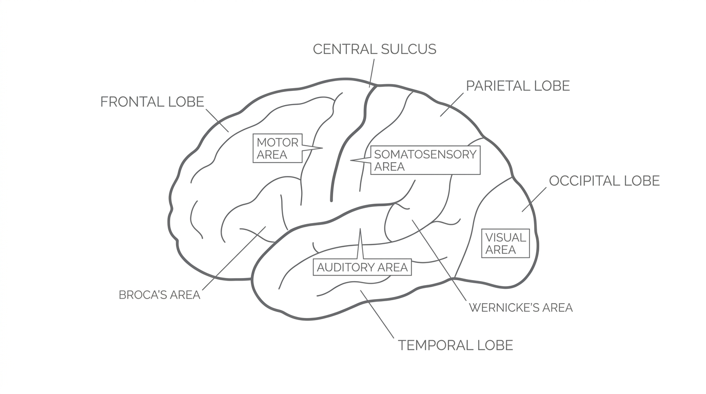
Part 2 — The Language Centres: Broca's and Wernicke's Areas
Unlike the motor, somatosensory, visual, and auditory centres — which appear in both hemispheres — the brain's language centres are found predominantly in the left hemisphere for most people. This makes language an important example of a function that is both localised and lateralised.
Broca's area
Broca's Area
A region of the left frontal lobe (Brodmann areas 44 and 45) responsible for speech production. Damage produces Broca's aphasia, characterised by slow, laboured, non-fluent speech.
In the 1860s, the French surgeon Paul Broca identified a small region of the left frontal lobe that appeared essential for producing speech. His most famous patient was Louis Victor Leborgne, nicknamed "Tan" because "tan" was the only syllable he could utter. A post-mortem examination of Tan's brain revealed a lesion in this left frontal area, which Broca linked to the patient's inability to produce fluent speech.
Damage to Broca's area results in Broca's aphasia: speech becomes slow, effortful, and halting. Patients typically produce only short, fragmented sentences and struggle with grammatical words such as prepositions and conjunctions (e.g. "a", "the", "and"). Crucially, however, their comprehension of language usually remains relatively intact — they understand what is said to them but cannot express themselves fluently.
Wernicke's area
Wernicke's Area
A region of the left temporal lobe (encircling the auditory cortex) responsible for language comprehension. Damage produces Wernicke's aphasia, characterised by fluent but meaningless speech containing neologisms.
Around the same period, the German neurologist Karl Wernicke described patients who had the opposite pattern: they could produce fluent, well-articulated speech, but what they said made little sense, and they had severe difficulty understanding language. Wernicke identified a region in the left temporal lobe — now called Wernicke's area — as being responsible for language comprehension.
Damage to Wernicke's area causes Wernicke's aphasia: the patient speaks fluently and with normal intonation, but their speech is littered with neologisms (made-up or nonsensical words). A hallmark of Wernicke's aphasia is that patients are typically unaware that their speech is meaningless — they do not recognise their own errors.
The contrast between the two aphasias provides powerful evidence for localisation: two distinct brain regions, both involved in language, produce entirely different patterns of impairment when damaged.
David, a 62-year-old retired teacher, suffered a stroke that damaged a region of his left frontal lobe. Since the stroke, David can understand everything his family says to him and follows conversations without difficulty. However, when he tries to reply, he produces only short, fragmented phrases — for example, saying "shop... bread... tomorrow" instead of "I need to go to the shop to buy bread tomorrow." David becomes visibly frustrated because he knows what he wants to say but cannot form the words fluently.
This pattern of preserved comprehension with impaired speech production is characteristic of damage to Broca's area and illustrates why precise knowledge of localised brain regions matters — it allows clinicians to predict which function will be affected based on the location of brain damage.
Part 3 — Hemispheric Lateralisation
Hemispheric Lateralisation
The idea that the two hemispheres of the brain are functionally different, with certain mental processes and behaviours being predominantly controlled by one hemisphere rather than the other. Language, for example, is lateralised to the left hemisphere in most people.
The brain's two hemispheres are broadly symmetrical in structure, but they are not identical in function. Some abilities — like language — are dominated by one hemisphere, a principle called lateralisation. Other functions, such as vision and motor control, are represented in both hemispheres but are contralaterally wired: the right hemisphere controls the left side of the body, and the left hemisphere controls the right side.
Left hemisphere — the "analyser"
The left hemisphere (LH) houses the main language centres (Broca's area and Wernicke's area) and tends to dominate in tasks requiring analytical processing, logical reasoning, and the sequential handling of information. Research with right-handed individuals shows that approximately 95% have left-hemisphere dominance for language.
Right hemisphere — the "synthesiser"
The right hemisphere (RH) is associated with visuospatial processing — the ability to perceive spatial relationships, recognise faces, process emotions, and handle tasks requiring holistic rather than piecemeal analysis. Although the RH can produce only rudimentary words and phrases, it contributes emotional tone and context to communication.
Contralateral organisation of vision
The visual system provides a clear example of contralateral wiring. Each eye receives light from both the left visual field (LVF) and the right visual field (RVF). Information from the LVF of both eyes travels to the right hemisphere's visual cortex, and information from the RVF of both eyes travels to the left hemisphere's visual cortex. In a normally connected brain, the corpus callosum immediately shares information between hemispheres, giving a unified picture. In a split brain, this sharing cannot occur — which is why split-brain research has been so informative.
Part 4 — Split-Brain Research: Sperry and Gazzaniga
Split-Brain Research
A programme of studies, beginning in the 1960s, involving patients who had undergone surgical severing of the corpus callosum (a commissurotomy) to control severe epilepsy. By preventing communication between hemispheres, researchers could test the functions of each hemisphere in isolation.
Corpus Callosum
The thick bundle of approximately 200 million nerve fibres connecting the left and right hemispheres. It enables the two hemispheres to communicate and share information. Severing it produces a "split brain."
The most important research into hemispheric lateralisation came from the work of Roger Sperry and Michael Gazzaniga. Their participants were individuals with severe epilepsy who had undergone a commissurotomy — surgical severing of the corpus callosum — to prevent electrical seizure activity spreading from one hemisphere to the other. This created a unique opportunity: because the two hemispheres could no longer communicate, researchers could present information to one hemisphere at a time and observe how each hemisphere responded independently.
Sperry (1968) — Studied eleven patients who had undergone split-brain surgery, using a specially designed apparatus. Each participant had one eye covered and images were flashed onto a screen for one-tenth of a second — too brief for the eyes to move and expose the image to both visual fields. This ensured that stimuli presented to the right visual field (RVF) were processed exclusively by the left hemisphere, and stimuli presented to the left visual field (LVF) were processed exclusively by the right hemisphere. This was a laboratory experiment conducted under strictly controlled conditions.
Key findings
-
Describing what you see: When an image was presented to the RVF (left hemisphere), the participant could verbally describe what they saw. When the same image was presented to the LVF (right hemisphere), the participant said they saw "nothing" or just "a flash of light." This is because the right hemisphere lacks the language centres needed for verbal description.
-
Left-hand recognition: Although participants could not verbally name objects presented to the LVF, they could use their left hand (controlled by the right hemisphere) to select a matching object from a group hidden behind a screen. They could even select an object that was associated with the stimulus — for example, selecting an ashtray after being shown a picture of a cigarette. This demonstrated that the right hemisphere had processed and understood the image; it simply could not express this understanding in words.
-
Simultaneous presentation: When two different words were presented at the same time — one to each visual field — participants would say the word shown to the RVF (processed by the LH language centres) and simultaneously write or select the word shown to the LVF using their left hand (processed by the RH visuospatial centres).
-
Emotional responses: When an emotionally provocative image (such as a pin-up picture) was flashed to the LVF, participants showed an emotional reaction — for example, giggling or blushing — but reported that they had seen nothing or just a flash of light. This suggested that the right hemisphere could process the emotional content of an image even though it could not produce a verbal report.
-
Face matching: The right hemisphere appeared to dominate in tasks requiring participants to match a face to a stimulus, consistent with its role in visuospatial processing.
Conclusions
Sperry's findings provided direct evidence that the two hemispheres serve distinct functions. The left hemisphere is verbal — it can describe, name, and analyse. The right hemisphere is non-verbal but excels in spatial recognition, emotional processing, and drawing. In the intact brain, the corpus callosum integrates these contributions seamlessly, but when it is severed, each hemisphere operates independently, revealing its specialised capabilities.
Priya is a split-brain patient who underwent a commissurotomy five years ago. During a neuropsychological assessment, a picture of a key is flashed to her left visual field. When the researcher asks Priya what she saw, she says she saw nothing. However, when asked to reach behind a screen with her left hand and select the object from a group of items, she correctly picks up the key. Priya is confused about why she chose that object and cannot explain her choice verbally.
Priya's behaviour illustrates a core finding of split-brain research: the right hemisphere (which received the LVF image) recognised the key and guided her left hand, but because the right hemisphere lacks language centres, Priya could not produce a verbal description of what she had seen.
Part 5 — Supporting Evidence and the Holistic Challenge
Evidence from brain scans
Modern neuroimaging has provided further support for localisation theory. Petersen et al. (1988) used brain scans to demonstrate that Wernicke's area was active during a listening task, while Broca's area was active during a reading task — confirming the distinct roles of these two language centres in comprehension and production respectively.
Tulving et al. (1994) used PET scans to show that semantic memories (general knowledge) were recalled from the left prefrontal cortex, while episodic memories (personal experiences) were recalled from the right prefrontal cortex. This demonstrated that even within a single function — long-term memory — different types of processing are localised to different brain regions.
The case of Phineas Gage
One of the most famous case studies supporting localisation is that of Phineas Gage (1848). Gage was a 25-year-old railroad construction foreman who survived a catastrophic accident in which an iron tamping rod was propelled through his left cheek, behind his left eye, and out through the top of his skull, destroying much of his left prefrontal cortex. Remarkably, Gage survived the injury and could walk and speak almost immediately. However, those who knew him reported dramatic personality changes: previously calm, reliable, and well-mannered, Gage became impulsive, quick-tempered, and socially inappropriate — he was said to be "no longer Gage." Damasio et al. (1994) later used modern imaging techniques to reconstruct the path of the rod through Gage's skull, confirming that the damage involved the prefrontal cortex bilaterally, in areas associated with rational decision-making and emotional processing. This case study supports localisation because it links a specific brain region (the prefrontal cortex) to a specific set of functions (personality regulation, decision-making, and impulse control).
The holistic challenge — Lashley's equipotentiality
Not all evidence supports strict localisation. Karl Lashley (1950) removed between 10% and 50% of the cortex in rats that were learning to navigate a maze. He found that no single area of the cortex was more important than any other — the extent of cortical removal mattered, but its location did not. Lashley termed this equipotentiality: the idea that all parts of the cortex contribute equally to complex behaviours such as learning. This suggests that higher cognitive functions may be distributed more holistically across the brain rather than being confined to discrete regions.
More recently, Dick and Tremblay (2016) reviewed modern neuroimaging evidence and found that only about 2% of contemporary researchers believe language is entirely controlled by Broca's and Wernicke's areas alone. Brain imaging has revealed "language streams" distributed across the cortex, including regions in the right hemisphere and subcortical structures such as the thalamus. This suggests that while Broca's and Wernicke's areas are clearly important for language, the full picture is more holistic than early localisation theory proposed.
Localisation theory is well-supported for basic sensory and motor functions and for the broad distinction between Broca's area (speech production) and Wernicke's area (language comprehension). However, complex cognitive functions like learning and language may involve distributed networks across the brain, meaning strict localisation is likely an oversimplification.
Evaluation Bank (AO3)
Strength: Split-brain research provided strong scientific evidence for hemispheric lateralisation because it was conducted under highly controlled laboratory conditions. Sperry (1968) ensured that stimuli were flashed for only one-tenth of a second to prevent eye movement, and participants had one eye covered, meaning each stimulus was processed by only one hemisphere. This level of control allowed researchers to establish a clear causal link between the hemisphere that received the stimulus and the type of response produced (verbal vs. visuospatial). Furthermore, the findings have been supported by research on normally connected brains: Fink et al. (1996) used PET scans to show that the right hemisphere was more active when participants attended to global features of an image (such as a whole forest), while the left hemisphere dominated when attending to fine detail (such as individual trees). This convergence of evidence from both split-brain and intact-brain studies strengthens our confidence that hemispheric lateralisation is a genuine feature of brain organisation, not merely an artefact of the split-brain procedure.
Limitation: A significant limitation of split-brain research is the difficulty in generalising from these findings to the wider population. Sperry's eleven participants all had severe epilepsy and had been taking anti-epilepsy medication for extended periods before surgery, which may have caused neurological changes that affected their cognitive performance independently of the commissurotomy. Additionally, the control group used for comparison did not have epilepsy, meaning that any differences observed could have been caused by the epilepsy itself rather than the split brain — this is a confounding variable. The very small sample size (N = 11) further limits generalisability, as individual differences in the extent of the surgical lesion and pre-existing brain organisation may have influenced results. This relates to the broader issue of idiographic versus nomothetic approaches: the findings offer rich detail about a small, atypical group but may not reflect how lateralisation functions in the general population.
Limitation: Localisation theory may oversimplify the relationship between brain structure and function, representing a form of biological reductionism. While evidence from Broca, Wernicke, and Sperry demonstrates that certain areas are associated with particular functions, modern neuroimaging research challenges the idea that these functions are rigidly confined to specific regions. Dick and Tremblay (2016) found that language processing involves distributed networks extending well beyond Broca's and Wernicke's areas, including right-hemisphere regions and subcortical structures like the thalamus. Similarly, Lashley's (1950) work with rats demonstrated that no single cortical area was essential for learning a maze route, suggesting that complex cognitive functions are processed holistically. Furthermore, cases exist of left-handed individuals whose language is lateralised to the right hemisphere rather than the left, contradicting the prediction that language is always a left-hemisphere function. This suggests that strict localisation provides a useful starting framework for understanding brain function but cannot fully account for the flexibility and distributed nature of neural processing — a point that supports a more holistic view of brain organisation for higher cognitive functions.