8.8 - The heart and circulation
Follow blood through the heart, lungs and body, and explain how chamber walls, valves, the septum and major vessels make double circulation work.
8.8 — Trace the double circulation
The human circulation contains two linked loops. The pulmonary circulation connects heart and lungs. The systemic circulation connects heart and the rest of the body. Blood passes through the heart twice in one complete journey, so this is a double circulatory system.
The heart has four chambers. The two atria receive blood returning in veins; the two ventricles contract to send blood out through arteries. A muscular septum separates the right and left sides, preventing oxygenated and deoxygenated blood from mixing.
Follow one complete route, keeping every arrow in the direction of blood flow:
body-tissue capillaries
-> body veins
-> vena cava
-> right atrium
-> valve
-> right ventricle
-> valve
-> pulmonary artery
-> lung capillaries (carbon dioxide leaves; oxygen enters)
-> pulmonary veins
-> left atrium
-> valve
-> left ventricle
-> valve
-> aorta
-> body arteries
-> body-tissue capillaries
Names such as artery and vein describe direction relative to the heart, not oxygen concentration. The pulmonary artery is an artery because it carries blood away from the right ventricle, although that blood is deoxygenated. The pulmonary veins are veins because they return blood to the left atrium, although that blood is oxygenated.
Here is a useful worked classification. A vessel carrying blood from the heart to the lungs must be an artery because its direction is away from the heart; its destination shows that it carries deoxygenated blood to the lung exchange surface, so it is the pulmonary artery. Reversing the route identifies the oxygenated pulmonary veins returning from the lungs.
The coronary arteries branch from the aorta and deliver oxygen and glucose to the heart muscle itself. The heart is an active organ and needs its own blood supply; the blood inside its chambers is not a substitute for capillary delivery to its muscle cells.
[DIAGRAM: asset_name: 8.8 - Heart connections; asset_slug: edexcel-gcse-biology-8-6-8-8-heart-double-circulation-route; recommended_method: image_gen; description: Portrait connection schematic of the four-chambered heart with large numbered callouts. Anatomical right is viewer left. 1 right atrium; 2 right ventricle; 3 left atrium; 4 left ventricle with thicker muscular wall; 5 grouped venae cavae enter right atrium; 6 pulmonary artery leaves right ventricle; 7 grouped pulmonary veins enter left atrium; 8 aorta leaves left ventricle; 9 example atrioventricular valve, with outlet valves also drawn; 10 septum. Blue inward/outward arrows mark deoxygenated inflow/outflow, red arrows oxygenated inflow/outflow. Independent inlet-atrium-valve-ventricle-outlet-valve-artery paths, no mixing or crossing. Tube positions are schematic, not anatomically exact or to scale; use the normal-text numbered key below.]
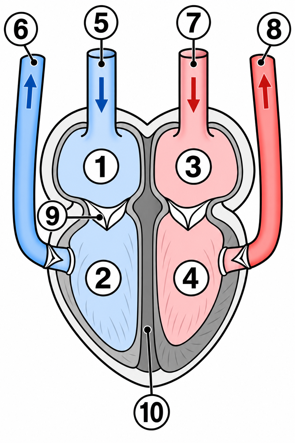
Read the numbered heart diagram
This is a simplified connection diagram, not a scale drawing of vessel positions. The heart’s right side is on your left. Blue represents deoxygenated blood and red represents oxygenated blood; deoxygenated blood is not actually blue.
Continue this lesson.
You’re previewing NovaLearn without an account. Create a free account to read the rest.
Create free account