8.7 - Blood vessels
Compare arteries, veins and capillaries, explaining how their walls, lumina and valves suit the pressure, direction and exchange demands of blood flow.
8.7 — Vessels are built for different pressures
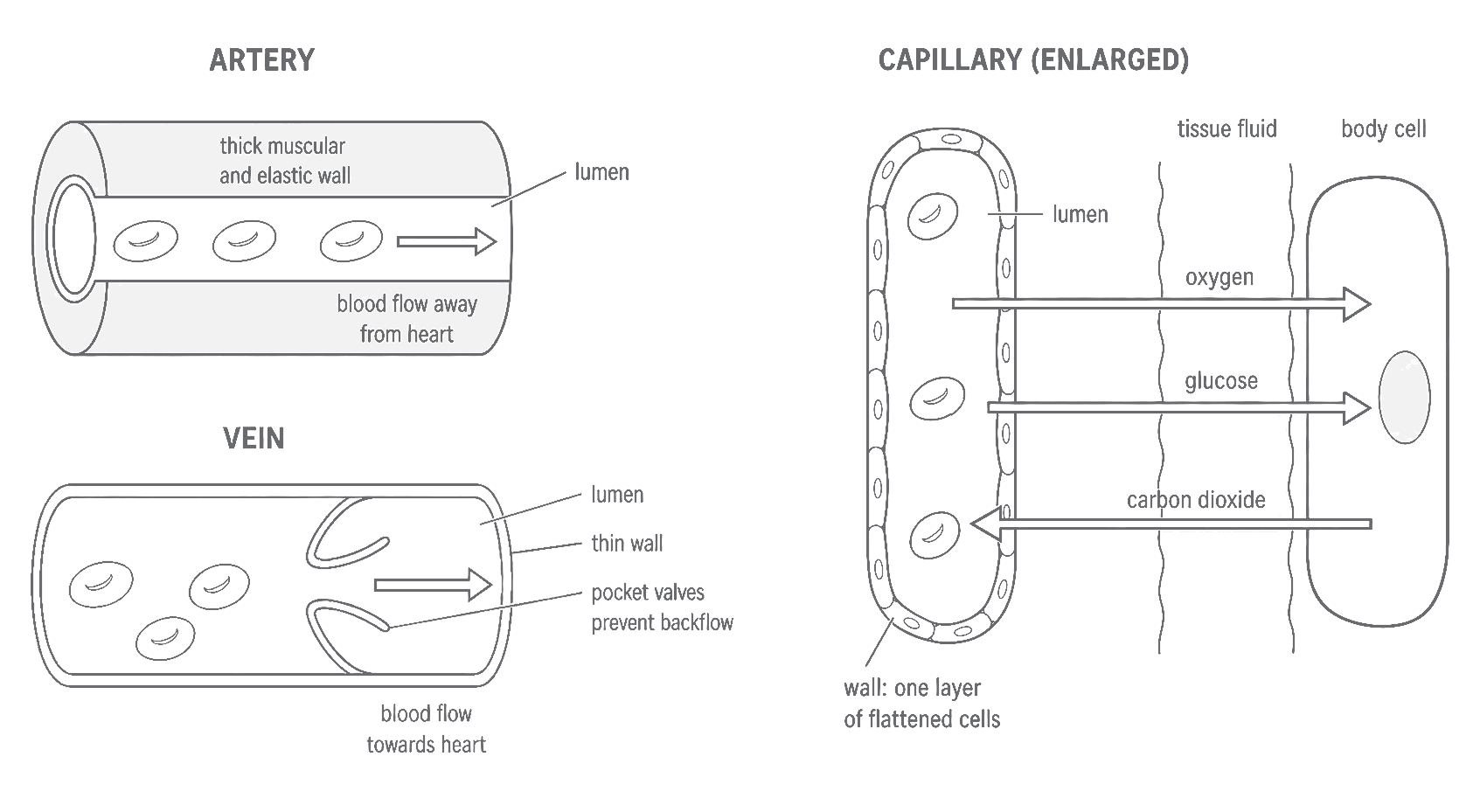
A vessel's lumen is the space through which blood flows. The three main vessel types form a route: arteries branch into smaller vessels and capillaries, while capillaries join into veins.
An artery carries blood away from the heart. Blood has just been forced out by a ventricle, so arterial pressure is relatively high and pulses with each contraction. Arteries therefore have thick, strong walls containing smooth muscle and elastic fibres. The wall withstands the pressure; elastic fibres stretch as blood enters and recoil between contractions, helping maintain forward flow.
A vein carries blood towards the heart. By the time blood has crossed a capillary network, its pressure is much lower. Veins can therefore have thinner walls with less muscle and elastic tissue, and a wide lumen offers little resistance to the returning blood. Many veins, especially in the limbs, contain valves. When surrounding skeletal muscles squeeze a vein, blood is pushed along; the valves close if it begins to move backwards, so the net direction remains towards the heart.
A capillary links the smallest arterial branches to the smallest venous branches. A capillary wall is only one flattened endothelial cell thick, giving a short diffusion distance. Networks of many capillaries provide a large exchange area, and their narrow lumina keep blood close to the wall.
[DIAGRAM: asset_name: 8.6-8.8 - Blood, vessels and the heart - diagram 01; asset_slug: edexcel-gcse-biology-8-6-8-8-vessel-structure-exchange; recommended_method: image_gen; description: Simple monochrome textbook comparison of an artery, a vein and a capillary. Show and label each lumen; the artery's thick muscular and elastic wall with an unmistakable flow arrow away from the heart; the vein's thinner wall, wider lumen and two pocket valves with an unmistakable flow arrow towards the heart; and a capillary wall consisting of one layer of flattened cells. Add a small body-cell exchange inset beside the capillary with oxygen and glucose arrows from the blood across the capillary wall to tissue fluid and a body cell, and a carbon dioxide arrow from the body cell through tissue fluid into the blood. Keep red blood cells inside the lumen, use no colour coding, show no numerical pressure values, and exclude advanced histological layer names.]
Continue this lesson.
You’re previewing NovaLearn without an account. Create a free account to read the rest.
Create free account