5.22B - Using monoclonal antibodies
Higher tier, separate Biology. Apply specific antibody binding to pregnancy tests, scans that locate clots or cancer cells, and targeted cancer treatment. Explain both the useful selectivity and its limitations.
5.22B · Detecting hCG in a pregnancy test
A monoclonal antibody is one of many identical antibodies from a single cell clone. Its binding site fits a particular target, so it can carry a detectable label to that target.
A pregnancy test does not detect a baby directly. It detects the hormone human chorionic gonadotrophin (hCG) in urine. The hCG acts as the antigen target for monoclonal antibodies with complementary binding sites.
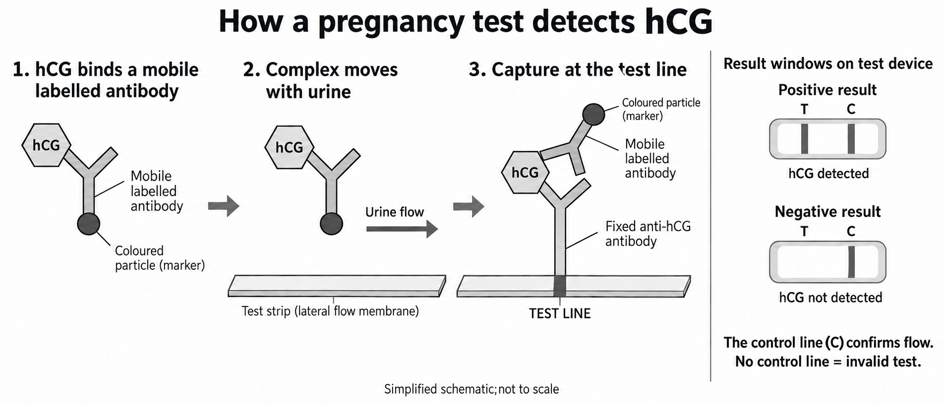
Inside a typical test strip, some anti-hCG monoclonal antibodies can move and have coloured particles attached to them. Other anti-hCG antibodies are fixed at the test line. They bind a different accessible site on the same hCG molecule, so hCG bridges the mobile antibody and the fixed capture antibody. These mobile and fixed antibodies have different jobs:
- Urine moves along the strip by capillary action.
- If hCG is present, it binds to the complementary mobile antibodies carrying coloured particles.
- The hCG-antibody complexes continue moving until fixed antibodies at the test line capture them.
- Many coloured particles accumulate in the same narrow region, making the test line visible.
A separate control line confirms that liquid and labelled antibodies moved through the strip. In a correctly manufactured, correctly used test, a control line by itself is read as a negative result (hCG not detected); a test line together with a valid control indicates that hCG was detected. The control line does not detect hCG and cannot replace the test line. No control line means the test is invalid. A control line confirms flow, but it cannot rule out every fault, such as missing capture antibodies at the test line.
[DIAGRAM: asset_name: 5.21B-5.22B - Monoclonal antibodies - diagram 01; asset_slug: edexcel-gcse-biology-5-22b-pregnancy-test-flow; recommended_method: image_gen; description: Three-stage schematic: hCG binds a mobile colour-labelled anti-hCG antibody, the complex moves with urine, and a fixed anti-hCG antibody at the test line binds another site on the same hCG molecule. The two antibodies touch only through hCG; the coloured particle stays on the mobile antibody. Separate result windows show T and C lines for positive, C only for negative, and explain that no control line means invalid. Simplified and not to scale.]
5.22B · Locating clots and cancer cells
For diagnosis inside the body, a monoclonal antibody can be joined to a detectable marker, such as a radioactive substance. The antibody supplies the biological specificity; the marker supplies the signal that a scanner can detect.
The route has four causal steps:
- Choose a monoclonal antibody whose binding site is complementary to a target associated with the tissue or structure being sought.
- Attach a detectable marker to the antibody and introduce the labelled antibody into the body.
- The antibody circulates and binds where its target is present, so more marker becomes concentrated at that location than in surrounding tissue.
- A detector maps the marker signal, revealing the position of the target.
Continue this lesson.
You’re previewing NovaLearn without an account. Create a free account to read the rest.
Create free account