H567/02 Section A - Sperry (1968): split brain study
Sperry used carefully separated sensory inputs to investigate what the two hemispheres could do after their main connecting pathways had been cut. The striking finding was a difference between what a patient could say and what the same patient could demonstrate through a hand response.
Background, design and patients
The cerebral hemispheres normally exchange information through bundles of nerve fibres, especially the corpus callosum. Earlier clinical observations suggested that cutting these connections produced surprisingly little obvious change in everyday behaviour. Animal research nevertheless indicated that specialised tests could reveal losses of communication between the hemispheres. Sperry wanted to investigate the functional consequences in humans, including lateralisation and the apparent unity of conscious awareness.
Lateralisation
The relative specialisation of the left and right cerebral hemispheres for particular functions. It does not mean that ordinary people use only one hemisphere.
The report concerned 11 patients who had undergone commissurotomy to treat severe epilepsy that medication had failed to control. Their corpus callosum and other cerebral commissures had been divided. Surgery was a clinical treatment, not an operation assigned by Sperry for research. The first patients had been studied for years; more recent patients had shorter follow-up. Their histories and recovery were not identical.
This was intensive research on a small clinical series, using controlled laboratory tasks. It is commonly classified as a quasi-experiment because the relevant split-brain condition already existed and could not be randomly allocated. Comparisons also drew on expected intact-brain performance. Within patients, researchers could vary the visual field receiving a stimulus or the hand receiving an object. Calling it quasi-experimental does not mean that every task lacked manipulation: the sensory presentation was controlled, while the clinical condition was not assigned.
The dependent measures included verbal identification, selection of matching objects, drawings and performance on other sensory tasks. The original article summarised a programme of tests, rather than one uniform trial with a single numerical outcome for every patient.
Isolating visual and tactile input
With gaze fixed centrally, information in the left visual field initially reaches the right hemisphere, and information in the right visual field initially reaches the left hemisphere. This division concerns the part of the visual scene, not which eye is used. Each eye contributes to both visual fields.
Patients sat at an apparatus with an upright translucent screen. With one eye covered, they looked at a central fixation point. A projector with a shutter flashed pictures, words or symbols from 35-mm transparencies to one side of fixation for one tenth of a second or less. That brief presentation prevented an eye movement from bringing the stimulus into the other field. Fixation and exposure duration were therefore essential controls, not incidental apparatus details.
The apparatus also concealed the hands from view. Researchers could place an object in one hand and ask for its name, or allow that hand to find a matching object among alternatives. Touch from the right hand is primarily processed by the left hemisphere; touch from the left hand by the right. Preventing sight of the object stopped visual information giving the other hemisphere the answer. Speech and other signals also had to be controlled to avoid indirect cueing.
[DIAGRAM: asset_slug: cs19_visual_response_routes; file: cs19_visual_response_routes.png; description: Brief left-field input reaches the right hemisphere and can support left-hand selection; right-field input reaches the left hemisphere and can support verbal naming. Severed commissural links prevent normal sharing.]
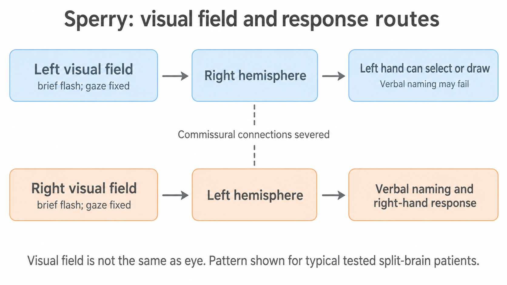
Diagram description: Follow each row from input to hemisphere to response. These are the typical routes demonstrated under the controlled tests; the diagram does not claim that all motor control is exclusively contralateral or that every patient had identical language organisation.
For example, a new picture flashed on the left could be recognised by pointing with the left hand even if the patient could not name it. Asking only for a spoken answer would miss that distinction. The choice of response tells us which capacities are accessible to the hemisphere that received the input.
Visual and tactile results
In the typical right-handed patients described, left-hemisphere input supported spoken and written identification. Input confined to the right hemisphere could support recognition through an appropriate nonverbal response, even when spoken identification failed.
| Controlled presentation | Typical demonstrated response | What the contrast shows |
|---|---|---|
| Picture in right visual field | Patient could describe/name it verbally | Left hemisphere had access to the visual information and expressive language |
| Picture in left visual field | Patient reported nothing identifiable or a flash, yet could select the matching item with the left hand | Failure to say the name was not proof that the right hemisphere had failed to perceive it |
| Dollar sign in left field and question mark in right field simultaneously | Left hand drew the dollar sign out of sight; spoken report identified the question mark | Different response systems could act on different input without normal sharing |
| Object placed in right hand out of sight | Patient could usually name it | Information from that hand reached the language-supporting hemisphere |
| Object placed in left hand out of sight | Verbal naming failed, but the left hand could retrieve the same object from a collection | Recognition and memory were available through a nonverbal route |
Objects recognised by touch with one hand could be retrieved again with that same hand, whereas cross-retrieval with the other hand failed under controlled conditions. Similarly, left-field visual input could guide left-hand selection, but not reliably right-hand selection. These tasks exposed difficulty transferring information, rather than a simple loss of touch or vision.
The right hemisphere also showed capacities such as selecting related objects and performing some simple language-comprehension tasks nonverbally. It was therefore too strong to conclude that it had no understanding of language. Expressive speech and comprehension are different claims.
Sperry reported important patient differences and exceptions. The table describes the central pattern, not a guarantee for every patient in every task. Some apparent deficits could also be compensated for through eye movements or indirect signals outside the test situation.
Continue this lesson.
You’re previewing NovaLearn without an account. Create a free account to read the rest.
Create free account