2.16B - 2.17B - Vision defects and correction
Distinguish focusing errors, cataracts and colour vision deficiency by where the fault occurs. Explain how lenses, corneal reshaping and lens replacement correct the appropriate defects. This Biology-only lesson applies to both tiers.
2.16B — When light misses the retina
The cornea and lens refract light; rod and cone receptors in the retina detect it. Rods support dim-light vision, while cones provide colour information. A fault can therefore affect focusing, the clarity of the lens, or receptor signals.
For a sharp view, light rays must be brought to a focus on the retina. A refractive defect occurs when the combined focusing power of the cornea and lens does not match the front-to-back length of the eyeball.
If rays meet too early, they cross and spread out again before reaching the retina. If they would meet too late, they are still converging when they reach the retina. Either way, each point on the object spreads over an area of the retina instead of producing a sharp focus, so vision is blurred.
| Defect | What is usually blurred? | Possible structural cause | What happens to the light? |
|---|---|---|---|
| Short-sightedness (myopia) | Distant objects; nearby objects are clearer. | The eyeball is too long, or the cornea/lens is too curved. | Light is refracted too much, so the rays focus in front of the retina. |
| Long-sightedness (hyperopia or hypermetropia) | Nearby objects; distant objects may be clearer, although stronger long-sightedness can blur both. | The eyeball is too short, or the cornea/lens is not curved enough. | Light is not refracted enough, so the rays would focus behind the retina. |
Be precise about the mechanism. "The eye is too big" or "too small" does not identify the important front-to-back length. Name that dimension and explain where the light rays meet. Saying that a sharp image forms in front of the retina, or would form behind it, is also valid optical wording; the result is a blurred pattern at the retina itself.
2.17B — Correcting refractive errors
A correcting lens changes the direction of light before it enters the eye. Its job is to compensate for the eye's excessive or insufficient refraction so that the cornea and lens together bring the rays to a focus on the retina.
| Defect | Correcting lens | Action on incoming rays | Final effect |
|---|---|---|---|
| Short-sightedness | Concave, also called a diverging lens; thinner in the middle | Spreads the rays out before they enter the eye, reducing the overall convergence. | Moves the focal point backwards from in front of the retina onto the retina. |
| Long-sightedness | Convex, also called a converging lens; thicker in the middle | Brings the rays together before they enter the eye, increasing the overall convergence. | Moves the focal point forwards from behind the retina onto the retina. |
[DIAGRAM: asset_name: 2.15B-2.17B - The eye and vision correction - diagram 02; asset_slug: edexcel-gcse-biology-2-15b-2-17b-refractive-defects-correction; recommended_method: image_gen; description: Monochrome textbook ray-path comparison with five clearly separated aligned panels: normal eye with light rays focusing on the retina; uncorrected short-sighted eye with rays focusing in front of the retina; short-sighted eye corrected by an external concave lens with rays focusing on the retina; uncorrected long-sighted eye with rays still converging at the retina and dashed construction lines meeting behind it; long-sighted eye corrected by an external convex lens with rays focusing on the retina. Label retina, cornea/lens, focal point, concave lens and convex lens; use unmistakable arrowheads and correct lens shapes; label the position where light rays focus rather than saying an image forms there; do not add numerical scales, clinical imagery, decorative colour or off-scope eye defects.]
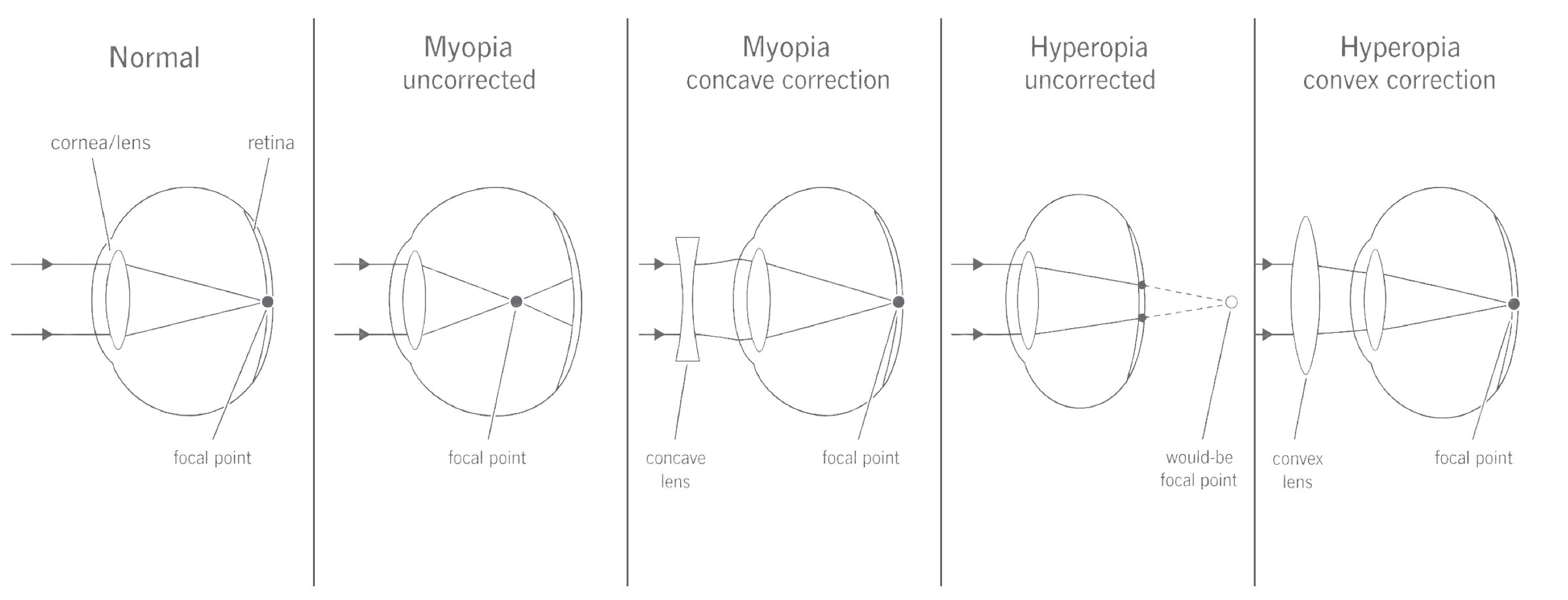
Prescription glasses and contact lenses both use this optical principle. They make vision clearer while they are being worn but do not shorten or lengthen the eyeball. For suitable patients, refractive laser surgery is another correction: it changes the curvature of the cornea so the eye refracts light by the amount needed to focus it on the retina.
2.16B–2.17B — Cataracts and colour blindness
Cataracts and colour blindness disrupt vision in different ways, and neither is the same kind of problem as short- or long-sightedness. A cataract affects the transparency of the lens; colour blindness affects receptor information from cone cells.
Cataracts
A cataract is a cloudy or opaque area in the normally transparent lens. Proteins in the lens can build up or clump, so light is scattered, dispersed or partly prevented from passing through. The retina therefore does not receive the sharply focused pattern of light it would receive through a clear lens, and vision becomes blurred or cloudy; colours can also appear faded.
Cataracts are corrected by surgery in which the cloudy natural lens is removed and replaced with a clear artificial lens. Simply saying "laser surgery" is not enough: opening or accessing the eye is not the correction. The key corrective step is replacing the cloudy lens.
Colour blindness
Colour blindness, more precisely called colour vision deficiency, occurs when one or more types of cone cell are absent or do not function normally. The brain then receives less distinct information about some wavelength ranges, making particular colours difficult to tell apart. Red-green difficulties are common, but colour blindness does not usually mean that a person sees only black and white.
This is a receptor defect, not a focusing defect. An ordinary convex or concave lens can change where light focuses, but it cannot restore a missing cone response. This is why the focusing lenses used for long- and short-sightedness are not the correction for inherited colour vision deficiency.
| First question to ask | Defect type | Appropriate response |
|---|---|---|
| Are rays focused at the wrong position? | Long- or short-sightedness | Change refraction with the correct converging or diverging lens; suitable corneal reshaping may also correct focus. |
| Is the natural lens cloudy? | Cataract | Replace it with a clear artificial lens. |
| Are cone signals for some colours reduced or absent? | Colour vision deficiency | Do not treat it as a front/behind-retina focusing error. |
Identify where the fault is before choosing a correction: ray focus, lens transparency and cone-cell information are three different biological problems.