2.13 - 2.14 - Nerves, synapses and reflex arcs
Trace how receptors detect a stimulus, neurones carry electrical impulses and synapses transfer the signal chemically. Use these structures to explain how a reflex moves a muscle rapidly without waiting for a conscious decision.
2.13 — From stimulus to electrical impulse
The nervous system allows the body to detect a change and make a response. A stimulus is a change in the internal or external environment, such as a rise in skin temperature, pressure on the skin, light entering the eye or a chemical in the air.
Sensory receptor
A specialised cell or nerve ending that detects a particular type of stimulus and converts it into an electrical signal.
Different receptors are sensitive to different kinds of stimulus. For example, temperature receptors in the skin respond to temperature change, while pressure receptors respond to mechanical force. This specificity means that the nervous system receives useful information about what has changed. A receptor can be the specialised ending of the sensory neurone itself, rather than always being a separate cell.
Detection and transmission are different jobs. The receptor detects the stimulus and starts the electrical signal. The sensory neurone then carries the electrical impulse from the receptor towards the central nervous system (CNS), which consists of the brain and spinal cord. An electrical impulse is a signal travelling along a neurone; it is not a flow of neurotransmitter along the whole cell.
2.13 — Neurones are built for direction
A neurone is a specialised cell that transmits electrical impulses. Its cell body contains the nucleus. The long, thin conducting parts allow an impulse to travel between structures that may be far apart in the body.
Direction matters when naming these parts:
- A dendron carries an electrical impulse towards the cell body. In a sensory neurone, the long dendron runs from the receptor to the cell body.
- An axon carries an electrical impulse away from the cell body. In a sensory neurone, a shorter axon continues from the cell body into the CNS; in a motor neurone, a long axon runs from the CNS to an effector.
- A myelin sheath forms an electrically insulating layer around much of a long conducting fibre. This insulation increases the speed at which electrical impulses travel.
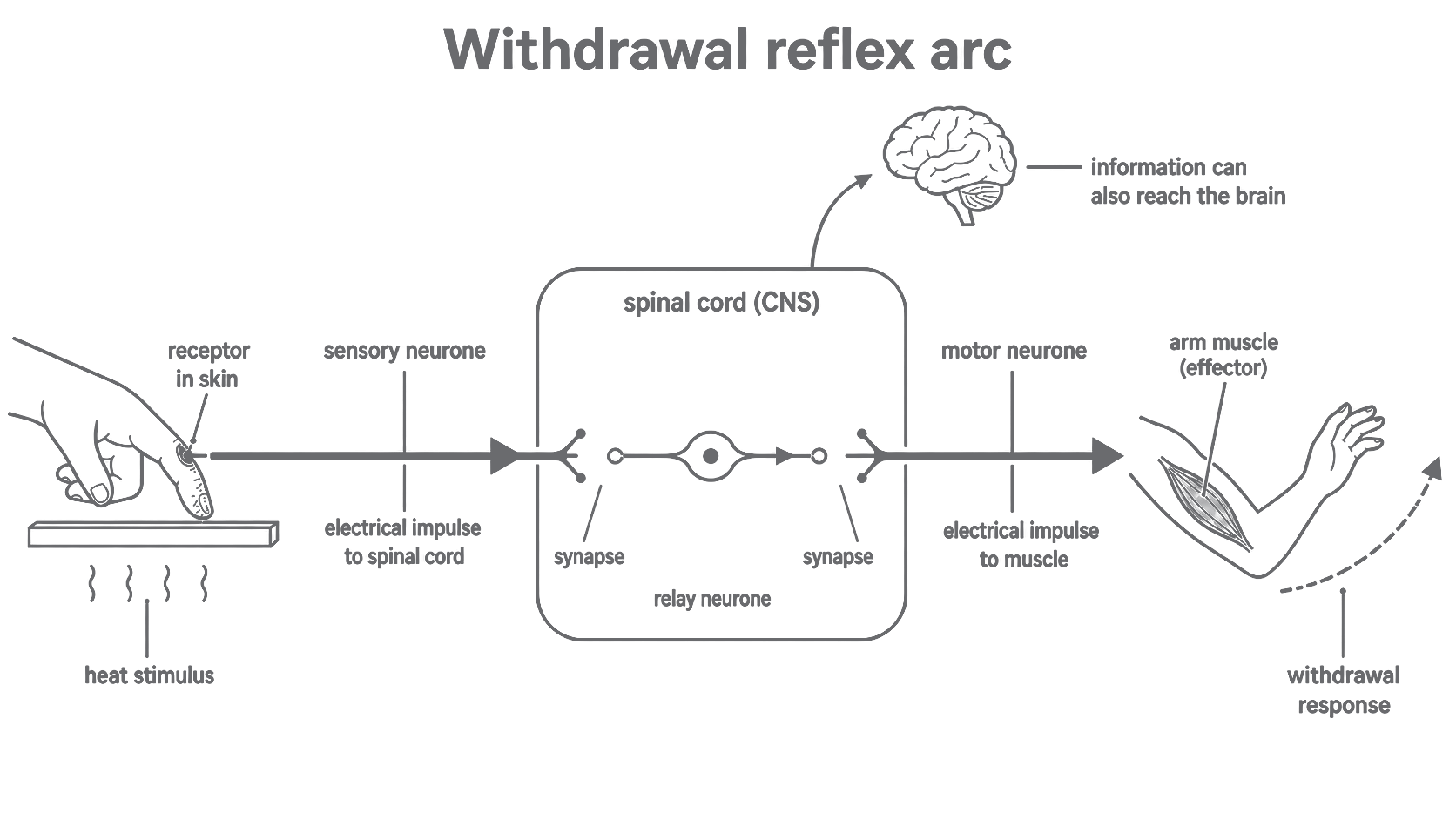
The three neurone types can now be distinguished by both route and structure.
| Neurone | Direction and function | Structure linked to function |
|---|---|---|
| Sensory neurone | Carries electrical impulses from a receptor towards the CNS. | A long dendron carries the impulse to a cell body positioned part-way along the route; a shorter axon carries it onwards into the CNS. The long conducting fibre is myelinated for faster transmission. |
| Relay neurone | Connects neurones within the CNS, for example linking a sensory neurone to a motor neurone. | It lies entirely in the brain or spinal cord and has short, branched processes suited to making connections with other neurones. |
| Motor neurone | Carries electrical impulses away from the CNS to an effector, such as a muscle or gland. | Its cell body and receiving branches are in the CNS, and its long myelinated axon carries the impulse to the effector. |
The comparison diagram simplifies the layout and is not to scale. In many skin sensory neurones the cell body sits on a short side branch of the conducting fibre; the important comparison is the receptor-to-CNS route and its dendron and axon, rather than a universal cell-body shape.
The myelin sheath does not create the impulse, and it does not carry a chemical message. Its structural contribution is insulation, with the consequence of faster electrical transmission along the neurone.
[DIAGRAM: asset_name: 2.13-2.14 - Nerves, synapses and reflex arcs - diagram 01; asset_slug: edexcel-gcse-biology-2-13-neurone-comparison; recommended_method: image_gen; description: Side-by-side simple textbook schematics of a sensory neurone, relay neurone and motor neurone with unmistakable impulse-direction arrows. Label receptor, long dendron, cell body, short axon and myelin sheath on the sensory neurone; show the relay neurone entirely inside a lightly outlined CNS region; label cell body, receiving branches, long axon, myelin sheath and effector on the motor neurone. Make relative layouts clear without implying exact scale. Exclude nodes of Ranvier, named glial cells, ion movements, photorealism and any structures beyond specification points 2.13-2.14.]
2.13 — A chemical handover at a synapse
Neurones do not form one continuous cell from receptor to effector. A synapse is the junction between a sending neurone and a receiving neurone, with a tiny gap between them. The signal must be transferred across this gap.
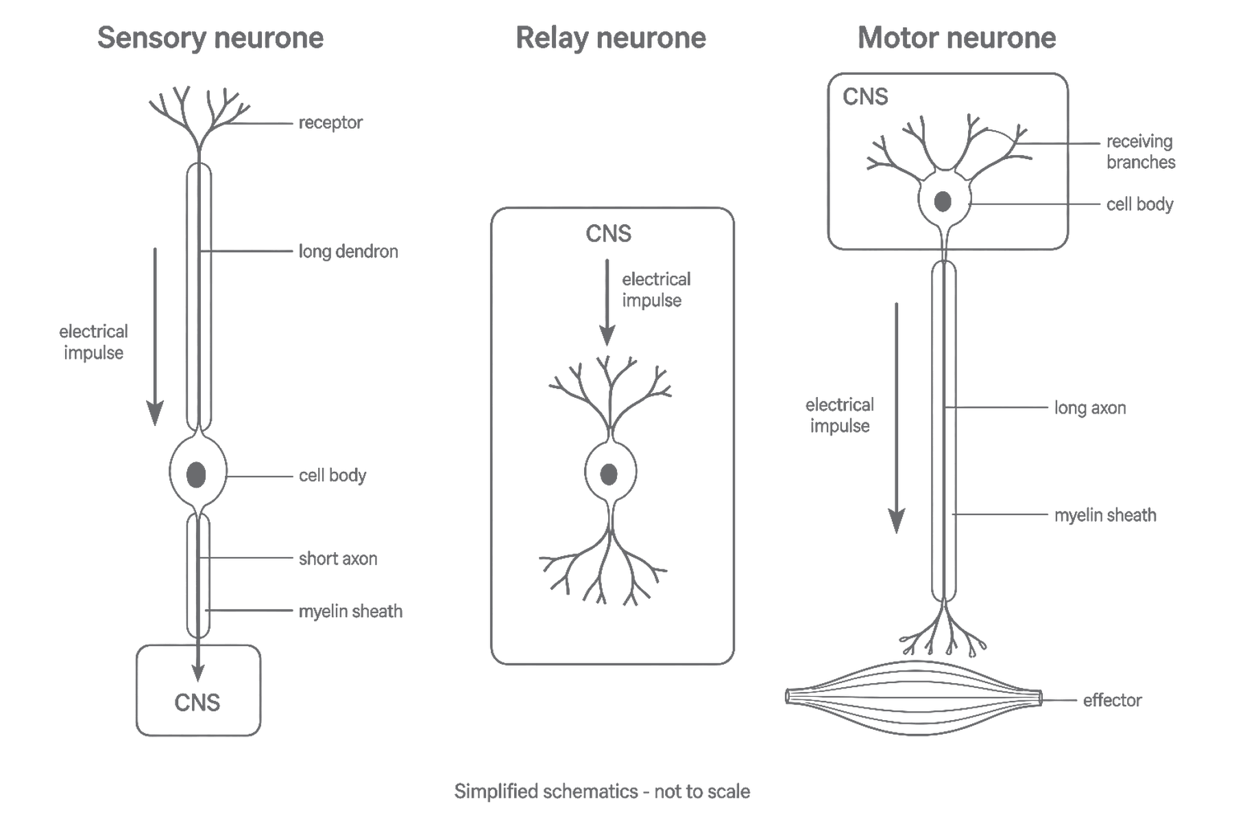
At the chemical synapses in this pathway, transmission occurs in a fixed sequence:
- An electrical impulse reaches the end of the axon of the sending neurone.
- Its arrival triggers the release of a chemical messenger called a neurotransmitter.
- The neurotransmitter diffuses across the synaptic gap and binds to receptor molecules on the membrane of the receiving neurone.
- This can trigger a new electrical impulse in the receiving neurone.
Release occurs at the sending ending and the matching receptor molecules are on the receiving membrane. This arrangement makes transmission through this synapse one-way.
The receptor molecules in step 3 respond to a neurotransmitter. They are not the sensory receptors that detect environmental stimuli at the beginning of a nervous pathway.
Electrical along a neurone -> chemical across a synapse -> electrical in the next neurone.
An electrical impulse therefore does not jump or diffuse across the synaptic gap. The impulse in the first neurone causes neurotransmitter release; the neurotransmitter is the substance that crosses the gap.
[DIAGRAM: asset_name: 2.13-2.14 - Nerves, synapses and reflex arcs - diagram 02; asset_slug: edexcel-gcse-biology-2-13-chemical-synapse; recommended_method: image_gen; description: Enlarged simple textbook schematic of one chemical synapse between a relay neurone axon ending on the left and a motor neurone membrane on the right. Show and label the incoming electrical impulse, neurotransmitter release, synaptic gap, neurotransmitter particles diffusing left-to-right, receptor molecules on the receiving membrane and the new electrical impulse in the motor neurone. Use arrows that distinguish movement along each neurone from diffusion across the gap. Exclude named neurotransmitters, vesicle proteins, ion channels, ion movements, numerical scales, photorealism and clinical content.]
2.13–2.14 — The protective reflex arc
Reflex
A rapid, automatic response to a stimulus that does not require a conscious decision.
A reflex arc is the nervous pathway that produces a reflex. Its value becomes clear when delaying a response could increase harm.
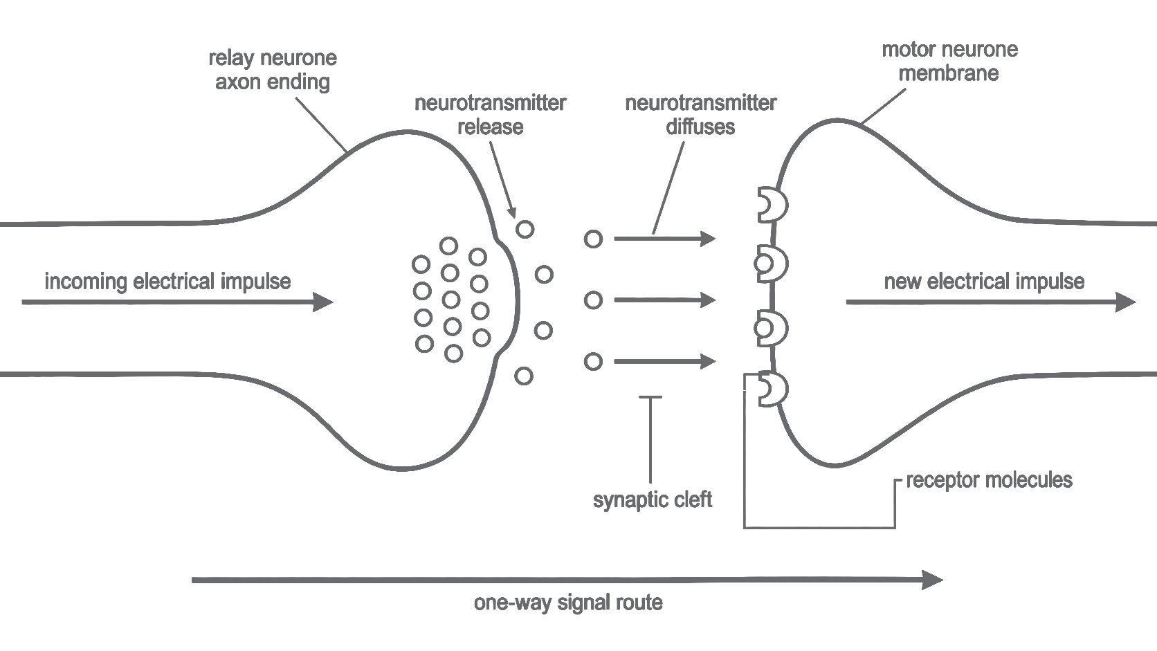
Worked route: withdrawing a hand from a hot tray
- Stimulus: the high temperature is a change that can damage tissue.
- Receptor: a temperature-sensitive or damage-sensitive receptor in the skin detects the stimulus and starts an electrical impulse.
- Sensory neurone: the impulse travels along the sensory neurone to the spinal cord, which is part of the CNS.
- Relay neurone: at a synapse, the incoming impulse triggers neurotransmitter release. The neurotransmitter diffuses across the gap and a new impulse begins in a relay neurone in the spinal cord. The same chemical handover at another synapse triggers an impulse in a motor neurone.
- Motor neurone: a new electrical impulse travels along the motor neurone from the spinal cord to an arm muscle.
- Effector and response: the muscle is the effector. It contracts, pulling the hand away and reducing the time for which the skin is exposed to the damaging heat.
The order of the named neurones is therefore sensory -> relay -> motor. The response is rapid because the spinal cord can coordinate it without waiting for a conscious decision by the brain. Information can still travel to the brain, so the heat and pain may be consciously perceived even though withdrawal has already begun.
[DIAGRAM: asset_name: 2.13-2.14 - Nerves, synapses and reflex arcs - diagram 03; asset_slug: edexcel-gcse-biology-2-14-withdrawal-reflex-arc; recommended_method: image_gen; description: Clear directional textbook schematic of a hand contacting a hot tray and withdrawing through the required reflex arc. Show and label stimulus, receptor in skin, sensory neurone carrying an electrical impulse into the spinal cord/CNS, relay neurone wholly inside the spinal cord, two synapses, motor neurone carrying an electrical impulse out to an arm muscle, muscle effector and withdrawal response. Arrowheads must give the single ordered route receptor -> sensory -> relay -> motor -> effector, with a separate small arrow from the spinal cord towards the brain labelled information can also reach the brain. Exclude detailed spinal-cord anatomy, antagonistic muscles, named neurotransmitters, reaction-time apparatus, photorealism and any suggestion that the motor neurone carries impulses towards the CNS.]